Chapter 2: Crime Scene Bloodstain Pattern Analysis
2.1: Basic Biological Properties of Human Blood
Blood is a bodily fluid circulating within the body.
An average adult has a blood volume of approximately 8% of his or her body weight.
Blood consists of a cellular portion as well as a liquid portion known as plasma.
Cellular Portion consists of blood cells and platelets.
Plasma is mostly composed of water and other substances such as proteins, inorganic salts, and other organic substances.
Blood can form clots (or thrombi) that are the result of blood coagulation.
Coagulation begins after an injury occurs, stopping blood loss from a damaged vessel.
The normal coagulation time for 1 mL of venous blood in a glass tube is 5–15 min.
The coagulation time can be affected by many factors such as blood volume and mechanical disturbance.
2.2: Formation of Bloodstains
Blood is viscous, and blood viscosity is a measure of the blood’s resistance to flow.
The viscosity of blood is approximately five times greater than that of water.
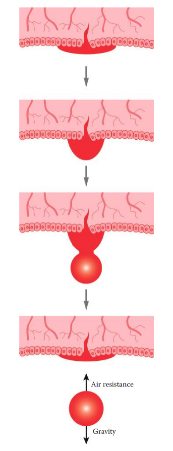
During the formation of a drop of blood, blood leaks out from a blood source.
The surface tension of the blood causes it to hang from the opening of a blood source and to form a pendant drop of blood.
The molecules of a blood drop are held together by the cohesion force to maintain the shape of a blood drop.
Surface tension causes liquids to minimize their surface, making the formed blood drop spherical.
As the volume of the drop gradually increases and exceeds a certain size, it detaches itself and falls. The falling drop is also held together by surface tension.
A falling blood drop is influenced by the downward force of gravity acting on the drop and the air resistance that acts in the opposite direction as the drop is in motion.
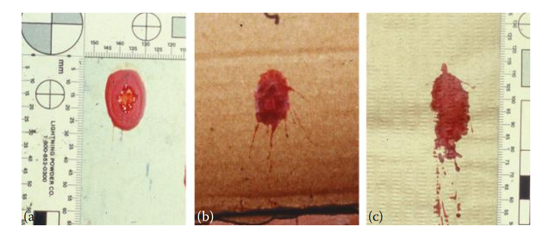
When a bloodstain lands on a surface, the shape and size of the bloodstain is affected by the texture of the target surface.
Bloodstains that land on porous or rough surfaces usually have more distortion around the edges of the stains than those that land on smooth surfaces.
2.3: Chemical Enhancement and Documentation of Bloodstain Evidence
Many chemical reagents react with blood to exhibit a color, a chemiluminescent light, or a fluorescent light. These tests are extremely sensitive and thus are used as chemical enhancement reagents for detecting bloodstains.
For bloodstain pattern analysis, the enhancement reagent is primarily used for detecting latent bloodstains such as diluted bloodstains that are visible on enhancement.
Luminol: Used for locating bloodstains at the scene.
Other reagents such as phenolphthalein, leucomalachite green, and tetramethylbenzidine are not often used as enhancement reagents but rather as presumptive tests for blood.
In bloodstain pattern analysis, special attention must obviously be given to bloodstains.
The photographic documentation of bloodstains may be performed by multiple means, including film and digital photography as well as videotaping.
Photographs should be taken with an overall view followed by a medium-range and a close-up view of the bloodstain patterns.
A scale of measurement must be included in the photograph, which is critical for bloodstain analysis.
To avoid any distortion, the photographs should be taken with the camera lens parallel to the target surface where the bloodstains are located.
An overall photograph provides an overall view of the scene including the bloodstain evidence.
A midrange photograph provides more details of the bloodstain pattern compared with that of the overall photograph.
Single bloodstains should be visible in midrange images.
A close-up photograph, usually taken with a macro lens, provides a detailed image of single bloodstains, which is useful for spatter pattern analysis.
2.4: Analyzing Spatter Stains
Spatter Stain: A bloodstain resulting from a blood drop dispersed through the air due to an external force applied to a source of liquid blood.
The patterns of spatter stains, including the shape and the size of the stains, are affected by the direction and the angle of impact of the spatter stains that are projected.
Velocity of Blood Droplets
Low Velocity Impact Spatter:
Formed when a blood droplet is traveling at <1.5 m/s.
The resulting stains are usually >4 mm in diameter.
As the traveling speed of blood droplets increases, the size of the spatter stain decreases.
Medium Velocity Impact Spatter:
Formed when in a blood source is subjected to a force associated with beatings or stabbings.
The resulting stains range from 1 to 4 mm in diameter.
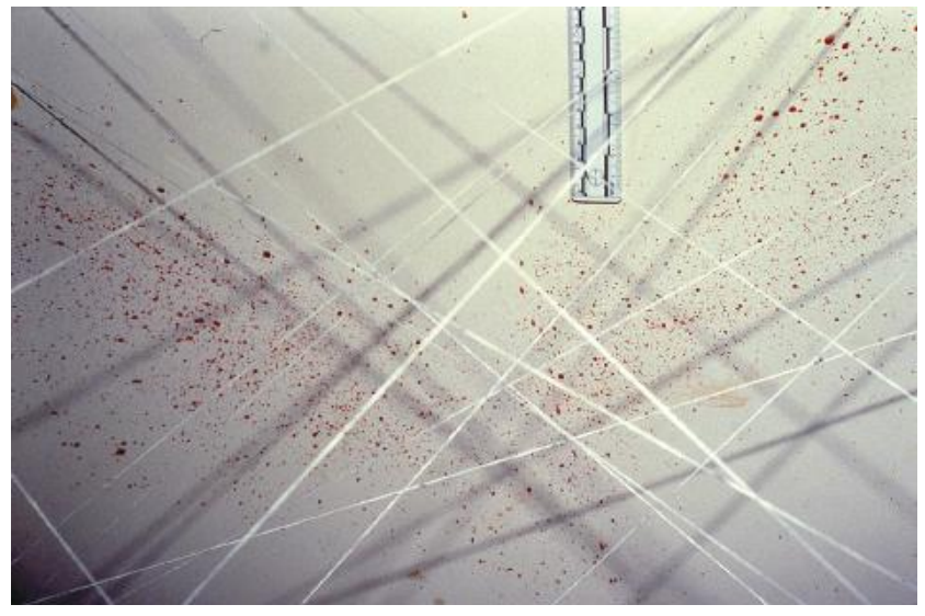
High Velocity Impact Spatter:
Formed when a blood source is subjected to a force associated with shooting using firearms.
The resulting stains are usually <1 mm in diameter.
Determining the Directionality of the Stains
Directionality: The characteristic of a bloodstain that indicates the direction blood was moving at the time of deposition.
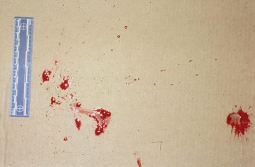
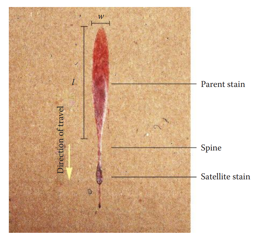
Parent Stain: The resulting spatter stain is an elongated ellipse.
Satellite Stain: A smaller bloodstain that originated during the formation of the parent stain as a result of blood impacting a surface.
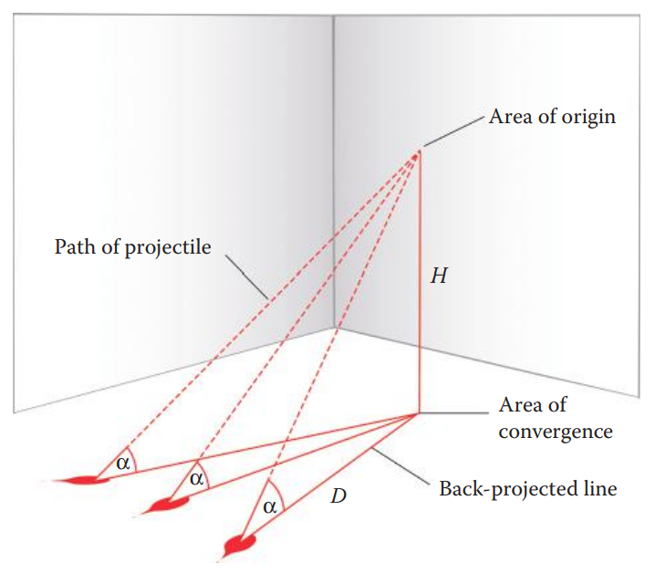
Determining Angles of Impact
Angles of Impact: The acute angle (alpha), relative to the plane of a target at which a blood drop strikes the target.
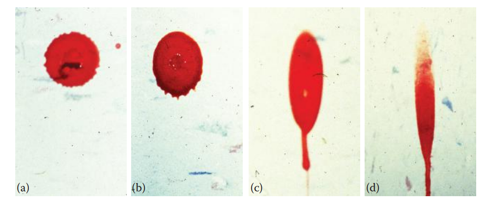
The shapes of the spatter stains are affected by the angle of impact.
When a blood drop lands on a surface at a perpendicular angle (90°), a circular parent stain is formed, where the length and the width of the stain are equal.
When a blood drop is projected onto a surface at an angle of between 0° and 90°, the stain is elongated.
As the impact angle decreases, the shape of the spatter stain is more elongated in which the length of the stain is greater than the width.
Determining Area of Origin
Area of Origin: The three-dimensional location from which spatter originated.
It can be determined based on the measurements from multiple elongated spatter stains
In the String Method, multiple well-formed, elongated spatter stains are selected for analysis.
A piece of string is then connected between the stain and a surface with one end of the string precisely attached to the spatter stain.
The path of the string, indicating the trajectory of the stain, is set using a protractor based on the calculated angle of impact.
In the Tangent Method, the directionality of a single stain is determined first.
A line is then back projected through the major axis of the bloodstain.
For a single impact event, approximately two dozen stains are processed to determine the area of convergence.
Area of Convergence: The area containing the intersections generated by lines drawn through the long axes of individual stains that indicates in two dimensions the location of the blood source.
2.5: Types of Bloodstain Patterns
Passive Bloodstains
It is formed due to bleeding from wounds, and the blood is deposited on a surface by the influence of the force of gravity alone.
Drip Stain: Formed when a falling drop of blood from an exposed wound or a blood-bearing object lands on a surface.
If a blood source is moving, a drip trail is formed.
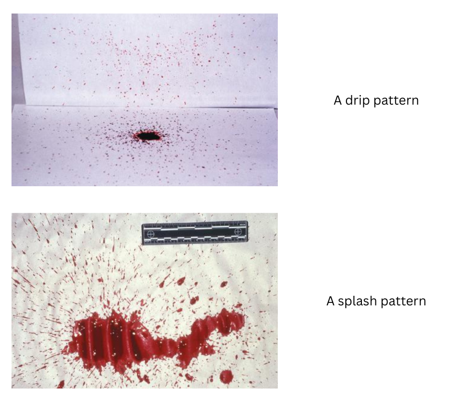
Drip Pattern: Formed when a liquid drips into another liquid, where one or both of the liquids are blood.
Secondary spatter stains are then generated.
As the dropping distance of the blood increases, the number of secondary spatter stains usually increases, and the size of these stains decreases.
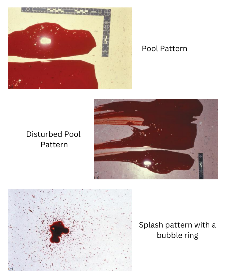
Splash Pattern: Formed when a volume of blood spills onto a surface.
It usually has large stains surrounded by numerous, peripheral, elongated bloodstains.
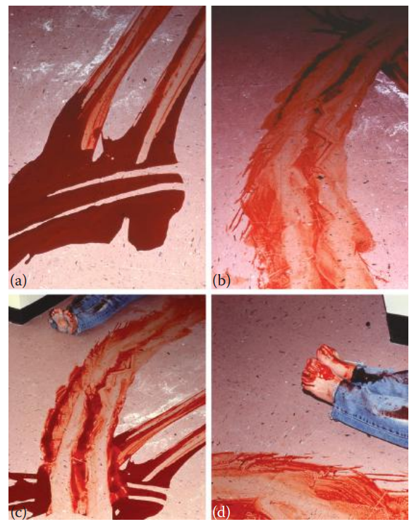
Flow Pattern: Caused by the movement of a large volume of blood on a surface either due to gravity or to the movement of the target such as a victim or postmortem disturbance.
Pool: A bloodstain resulting from the accumulation of liquid blood on a surface.
Sometimes, air bubbles in the blood may cause a bubble ring pattern.
If blood is coagulated, gelatinous blood clots can be observed.
Serum stain, which consists of the liquid portion of the blood after a clot is formed, may also be present.
Transfer Bloodstains
Transfer Bloodstain: A bloodstain resulting from contact between a blood-bearing surface and another surface.
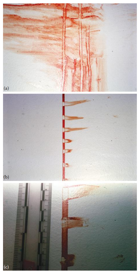
Swipe Pattern: A bloodstain pattern resulting from the transfer of blood from a blood-bearing surface onto another surface, with characteristics that indicate relative motion between the two surfaces.
Bloody impressions can provide information about the shape, the size, and the pattern of the objects such as finger ridges, hands, and shoe soles.
Wipe Pattern: An altered bloodstain pattern resulting from an object moving through a preexisting wet bloodstain.
Perimeter Stain: A type of wipe pattern, is a bloodstain that is disturbed before it is dried but it maintains the peripheral characteristics of the original stain.
Perimeter stain patterns can be useful for the estimation of sequential events of acts. The pattern can also be used to estimate a time frame between the time of bleeding and the subsequent act.
Projected Bloodstains
Projected Bloodstain: Formed when a volume of blood is deposited on a surface under pressure or a force that is greater than the force of gravity.
Impact Pattern: Formed when an object strikes liquid blood.
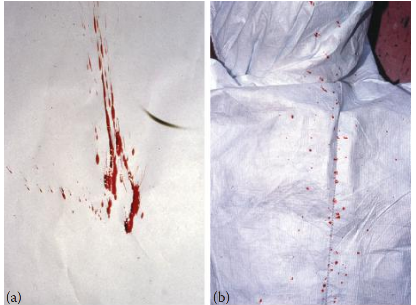
Cast-Off Pattern: Formed when blood drops are released from a moving blood-bearing object.
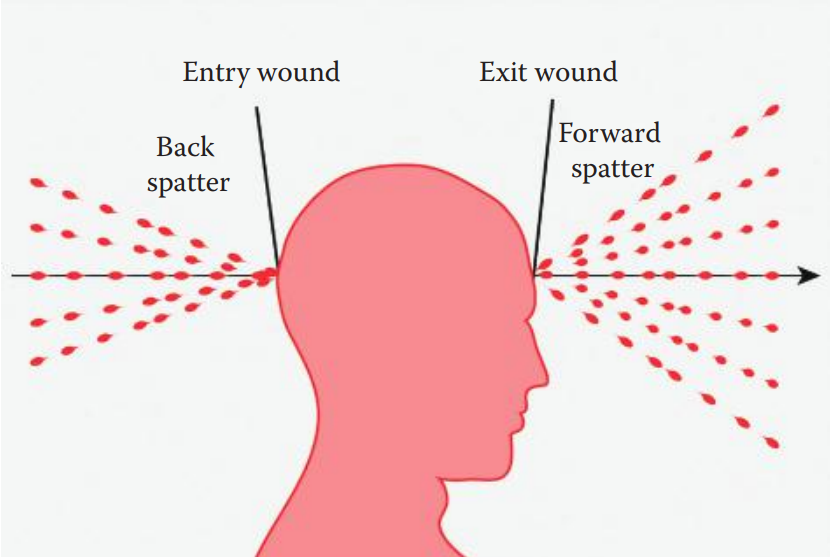
Forward Spatter: Formed when blood drops travel from an exit wound in the same direction as a projectile.
Back Spatter: Formed when blood drops travel from an entry wound in the opposite direction of a projectile.
Expiration Pattern: Formed when blood is forced by airflow through the trachea and out of the nose or mouth.
Arterial Spurt Pattern: Associated with wounds damaging arterial blood vessels where blood stains are driven by arterial pressure.
At a crime scene, if the projectile of bloodstains is blocked by an object, a void pattern is formed, which exhibits an area where there is an absence of blood surrounded by continuously distributed bloodstains.