Biochemistry
Study of chemical processes in cells
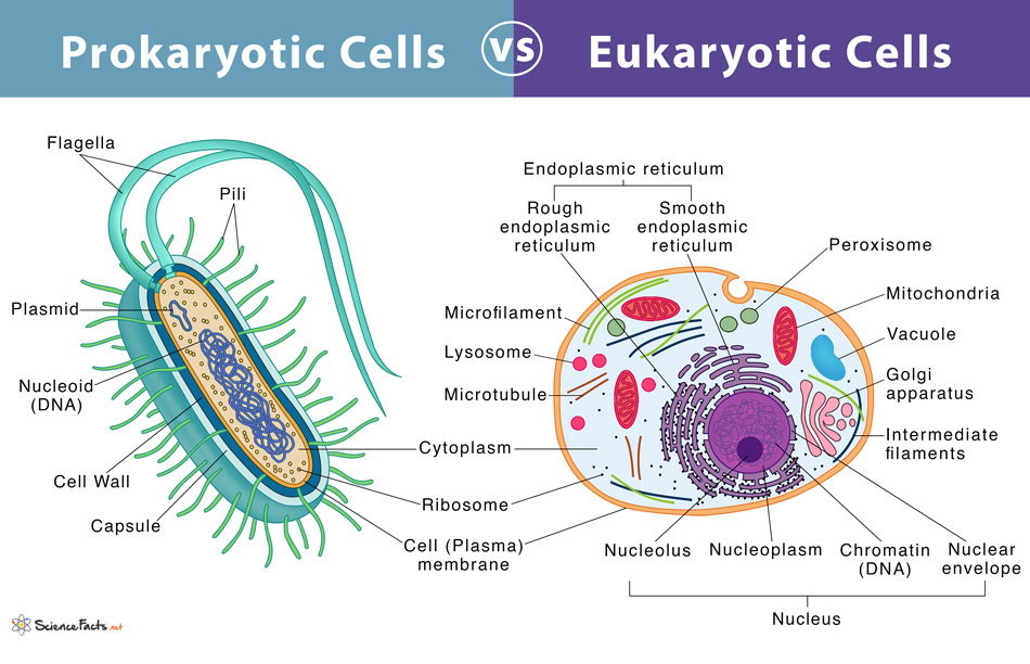
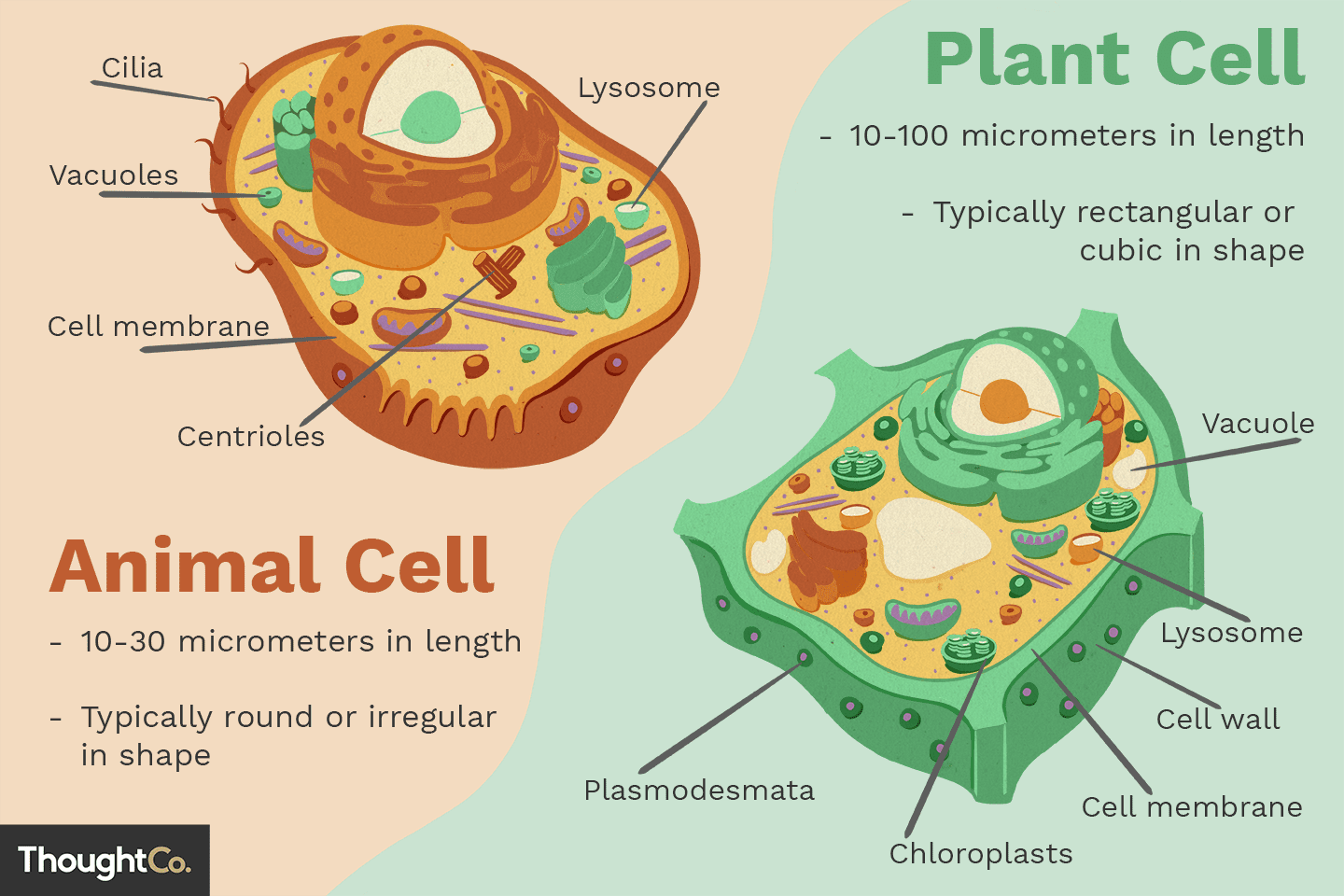
Prokaryotic Cells vs Eukaryotic Cells
Prokaryotic
Bacteria and archea
Membrane free nucleiod and structures
Eukaryotic
Fungi, animals and plants
Membrane bound nucleus and organelles
Organelle
Membrane enclosed structure in a cell
Cytosol
Fluid present in interior of the cell
Cell Membrane
Phospholipid bilayer with various protiens, enzymes, carbohydrates and lipids that encase a cell and organelles
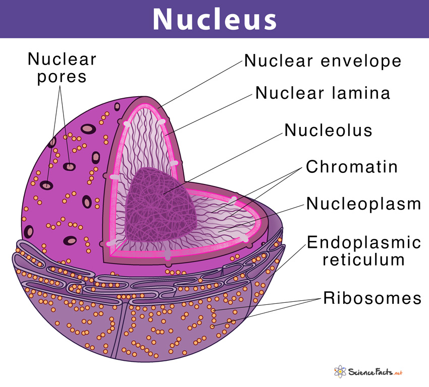
Nucleus
Location of DNA and directs protien synthesis
Nuclear Envelope
Double layered membrane sorrounding nucleus
Nuclear Lamina
Net-like layer of protiens that supports the shape of nucleur envelope from the inside
Chromosomes
Big coils of DNA wrapped around the protein histone
Nucleolus
Inner region of nucleos where RNA is transcribed
Nuclear structure
Ribosomes
Non membrane bound structure, made of rna and protiens, carries out protien synthesis
Free Ribsomes vs Bound Ribosomes
Free Ribosomes
Cytoplasm
Make protiens that function in cytosol
Bound Ribosomes
Held in membranes
Make protiens that function in membranes
Endomembrane System
Nucleus, ribosomes, endoplasmic reticulm, golgi apparatus, lysosomes, vesicles, vacuoles and plasma membrane.
Carries out protien synthesis, protien transport, metabolism and movement of lipids, and detoxing poisons
Vesicles
Sacks made of mebrane that transport protiens
Endoplasmic Reticulum
Folded membranes that sorround the nuclear envelope
Smooth ER
ER without ribosomes in membrane
Synthesis of lipids
Detoxing poisons
Metabolizing carbohydrates
Storing calcium ions
Rough ER
ER with ribosomes in the membrane
Creates glycoproteins in lumen(inner area)
Glycoproteins: Protein with carbohydrate attached
Transitional ER
Area of the ER used to transport proteins through transport vesicles
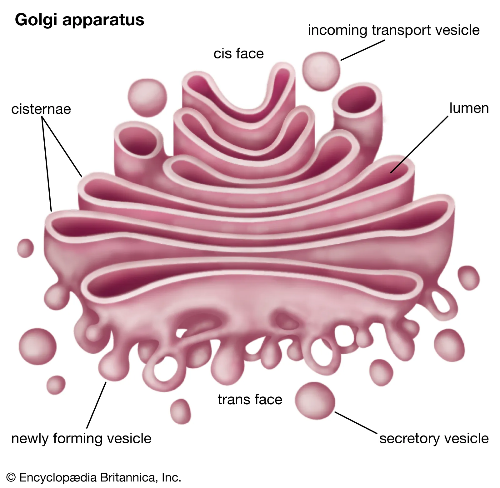
Golgi Apparatus
Processes, stores, and ships out products/protiens from ER. Made of many membrane sacks called cisternae. Finishes proteins from ER and creates non-protien products.
Cis face (Golgi Apparatus)
Recieves and absorbs product of the ER
Trans face (Golgi Apparatus)
Ships finished products out
Lysosome
Organelle containing hydrolytic enzymes to digest/hydrolyze macromolecules. Acidic interior. Made by ER and Golgi App
Phagocytosis
Unicellular protists eat by engulfing particles, forms food particle
Autophasy
Recycling products/materials in a cell
Vacuole
Large vesicle that serves as storage, different internal pH than cytosol. Carries out enzymatic hydrolysis in plants and fungi.
Contractile Vacuole
Pumps water from a cell
Central Vacuole
Large vacuole in mature plant cells, contains "cell sap" and inorganic ions. Grows and allows the cell to grow without new cytoplasm
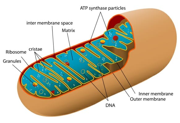
Mitochondria
Carries out cellular respiration. Found in almost all eukaryotic cells.
Mitochondria Structure
Contains its own ribosomes and DNA. Contains a folded inner membrane, folds called cristae
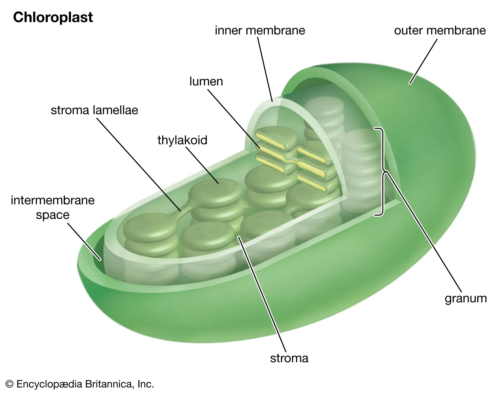
Chloroplasts
Cite of photosynthesis. Found only in plant cells. It is a plastid, a series of closely realted plant organelles including amyloplast and chromoplast.
Chloroplast Structure
Stroma is the inner liquid, like cytosol. It contains ribosomes and DNA. Thylacoids are stacked to form granules.
Peroxisomes
Single membrane bound metabolic compartment. Removes hydrogen atoms to create H2O2, then creates H2O. Breaks down fatty acids, detoxifys alcohol, etc
Glyoxysome
Peroxixomes found in fat storing tissue of plant seeds, used to make fatty acids into sugars
Cytoskeleton
The network of microtubules, centrosomes, centrioles, cilia, flagella, and microfilaments in a cell. Provides structure and anchors organelles.
Cell Motility
Movement of a cell and its organelles
Motor protiens
Guide organelles along microtubules of the cytoskeleton to transport them
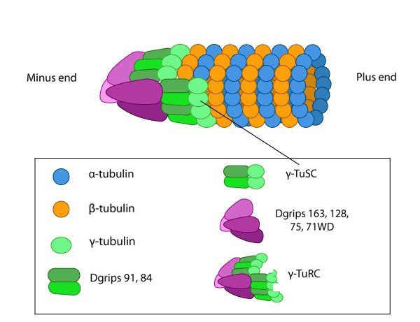
Microtubules
Compression resisting hollow rods made of the protien tubulins. Shape and support the cell, provide tracks for organelles to move.
Tubulins in microtubules
Tubulin dimers(joined alpha and beta), make up structure. The plus end of the structure is able to gain and lose tubulin more quickly than the other end.
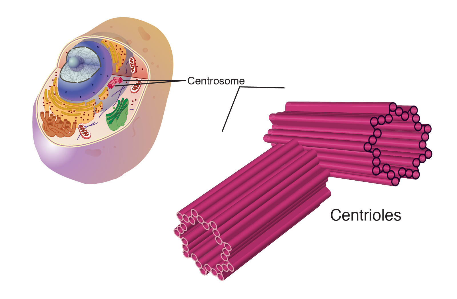
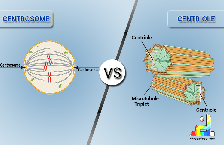
Centriole
9 sets of triplet microtubules in a ring shape.
Centrisome
Area near nucleous where most microtubules originate. Contains two centrioles(sometimes)
Basal body
Area at the base of microtubules that exit extracellular space. Structure of microtubules are covered by membrane and anchored to cell.
Dyneins
Protiens that cause the movement of cilia and flagella.
Flagella
Long, tail like structure that aids in movement of the cell
Cilia
Hair like structures that aid in movement of the cell
Primary cilia
Nonmotile, recieve signals for the cell
Internal structure of cilia and flagella
9+2 pattern, 9 doublets of microtubules wrapped around 2 single microtubules
Microfilament
Thin, solid rods made from actin(protien) twisted double chain. Thinner than microtubules. Tension bearing.
Cortical microfiliments
Just inside membranes, provide support to structure
Intermediate filaments
Bigger than microfiliments and smaller than microtubules. Found in some animal cells, diverse group made of keratin protiens. More permanent and structually supportive, make up nuclear lamina.
Microfilament v Microtubules
Microfilament
Actin double helix
Flexible but strong
Microtubules
Alpha and beta tubulin tubes
Stiff
Cortex
Outer cytoplasmic layer of cell
Myosin
Protien that interacts with microfiliments to contract muscle cells
Psuedopodia
Used by some cells for movement, myosin reliant
Cytoplasmic Streaming
Circular motion of cytoplasm in plant cells, allowing recources to be distributed. Reliant on microfiliments
Cell wall
Structure found in plant cells. Microfibrils made of cellulose join with other polysaccharides and protiens
Process of creating a cell wall
Primary, weak wall is made
Cell either strengthens original wall or creates second cell wall with a durable matrix
Middle lamella
Area between two cell walls of different cells. Contains pencin, which holds the walls together
Plant v animal cell
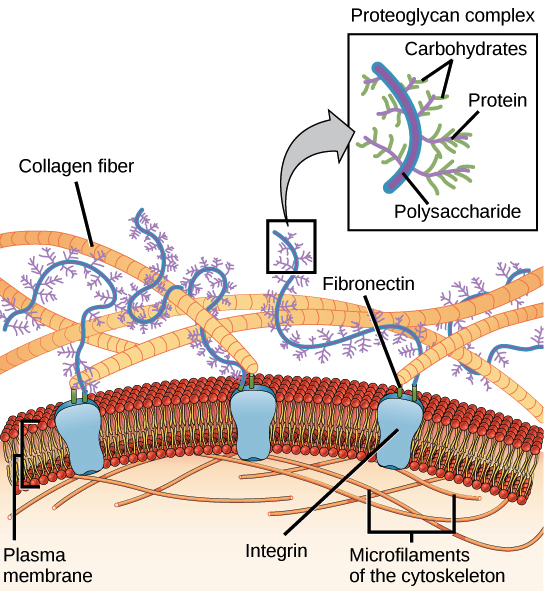
Extracellular matrix
Structures outside a cell that coordinate it's behavior. Constructed of glycoprotiens and carbohydrate containing molecules
Collagen
Protien found in extracellular matrix. 40% of protien in the human body
Proteoglycan
Protien core with carbohydrate chain
Fibronectin
Glycoprotien that bonds to integrins(receptor protiens)
Plasmodesmata
Channels that connect seperate plant cells
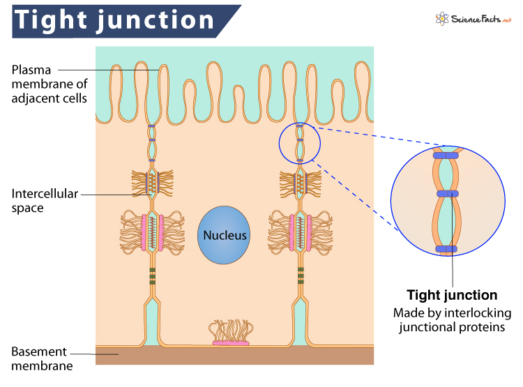
Tight junctions
Animal cells bond closely together, preventing anything from separating them
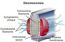
Desmosomes
Intermediate filaments pull to hold cells together, occurs with animal cells
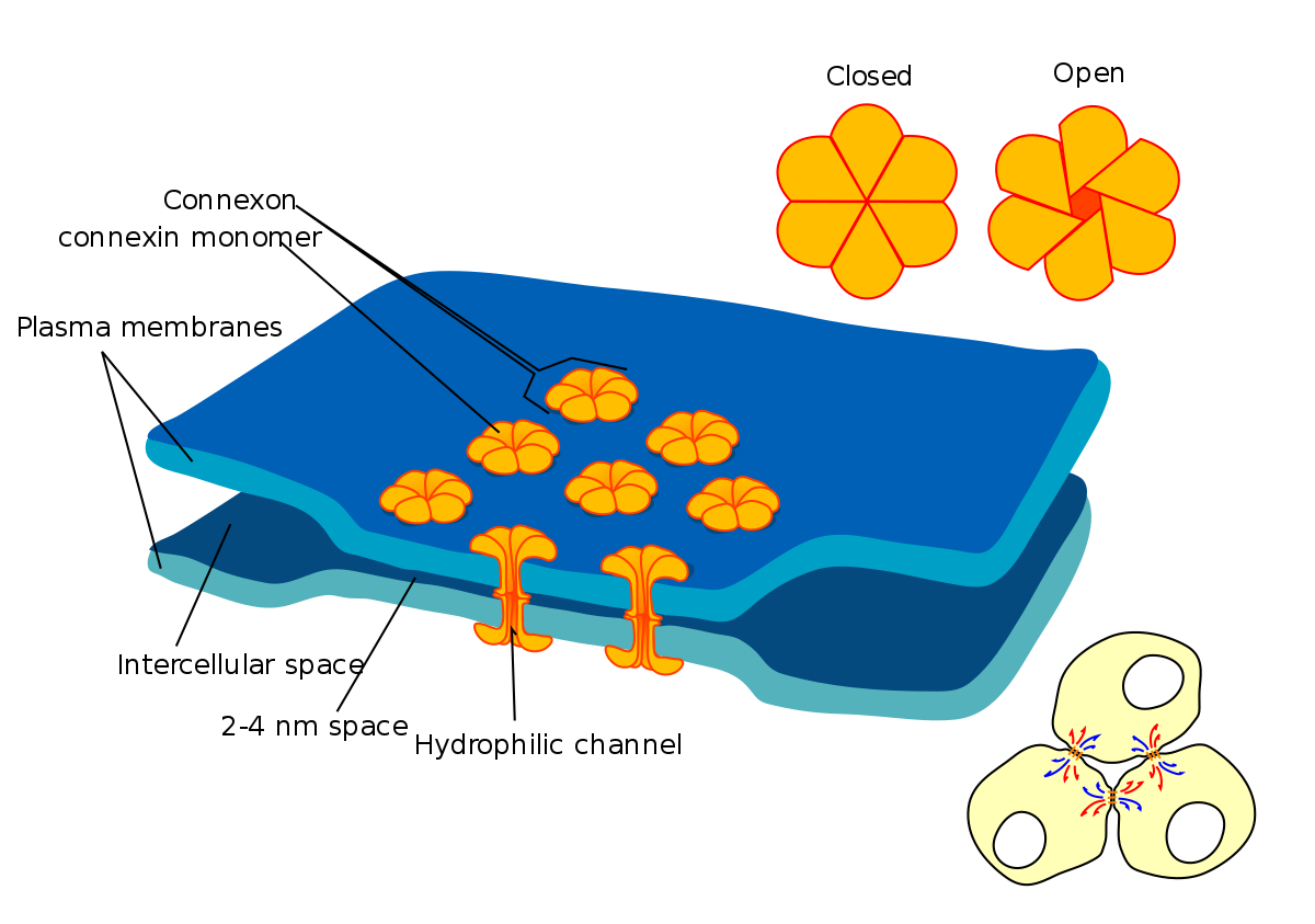
Gap junctions
Protiens from the membranes extend to create tunnels, occurs with animal cells
Amphipathic molecule
A molecule with a hydrophobic and hydrophillic end
Fluid mosiac model (membrane)
The modern theory that the cell membrane is full of constantly moving and fluid parts
What are some ways the membrane is fluid?
Held together by hydrophobic interactions
Lipids can fully flip around
Protiens can shift over time or be moved by cytoskeleton
Temperature's effect on membrane fluidity in animal and plant cells
Unsaturated(Plant)
Fluid, kinks are harder to compact
Less cholesterol Saturated(Animal)
Solid, no kinks are easier to compact
More cholestorol
Cholestorol in membranes
Acts as fluidity buffer, restrains movement but also makes it harder to pack
Interegal/transmembrane protiens
Protiens that extend the entire length of the membrane. Usually contain hydrophyllic passages
Peripheral protiens
Loosely bound to one side of the membrane. Could be attatched by cytoskeleton
Glycolipids
Carbohydrates bonded to lipids found in the membrane
Glysoprotiens
Carbohydrates attatched to protiens found in the membrane
Membrane carbohydrate function
Act as markers and communicators to other cells
Selective permiability
Membrane's ability to control what comes in and out. Hydrophobic molecules pass easily. Polar pass slowly and ionic have a harder time
Transport protiens
Allow hydrophillic and ionic molecules to pass through membranes easier.
Kinds of transport protiens
Channel - Create channel for hydrophillic molecules Aquaporins - Transfer water Carrier protiens - Forms around compounds and brings them through membrane
Passive transport
Diffusion of a molecule to create an equalibrium
Osmosis
Movement of free water to balance solution concentrations
Tonicity
Ability of sorrounding solution to affect a cell's water concentration
Isotonic
Equal solution inside and outside of cell, equal movement of water back and forth
Hypertonic
More solute and less free water outside cell, cell loses water
Hypotonic
Less solute and more free water outside cell, cell gains water
Osmosis reactions with cell wall
Isotonic - Flacid: Cell is flimsy and wilty Hypertonic - Plasmolyosis: Membrane shrivels away from cell wall, withers and dies Hypotonic - Turgid: Cell swells, stressing cell wall and creating stiffness
Osmosis reactions without cell wall
Isotonic - Normal Hypertonic - Shiveled Hypotonic - Lysed: Cell swells and bursts
Facilitated diffusion
Diffusion of polar and ionic molecules through transport protiens, form of passive diffusion
Active transport
Cell needs to expend energy because it moves against the concentration gradient. Powered by ATP hydrolysis
Membrane potential
Voltage across membrane, negative on cytoplamic side compared to extracellular side
Electrochemical gradient
The chemical and electrical forces acting on ions
Electrogenic pump
Transport protien that generates voltage
Proton pump
Electrogenic pump used in plants, pumps out H+
Cotransport
A transport protien uses the energy from brining in a molecule to push out another molecule
How large molecules leave the cell
vesicles
Exocytosis
The process by which large molecules are secreted from the membrane after being transported by vesicles
Endocytosis
The process by which large molecules are brought into the cell and transported by vesicles
Phagocytosis
Cell ingulfs a particle and metabolizes it
Pinocytosis
Cell takes in drops of extracellular fluid