Looks like no one added any tags here yet for you.
Information on the karyotype:
➢Size of chromosome
➢Position of centromere
➢Presence of secondary constrictions
➢Size of satellites
TRUE OR FALSE: Mutations cannot be detected in Karyotping
True
Karyotype
is the number and appearance of chromosome in the nucleus of a eukaryotic cell.
Karyotype comes from the Greek word…
Karyon
Karyon meaning
Nucleus
Karyology
the study of whole sets of chromosomes
Idiogram or Karyogram
the standard format of representing chromosomes as a diagram when the haploid set of chromosomes of an organism are ordered in a series of decreasing size.
Procedure
Karyotyping
The representation/visualization of result of karyotyping
Idiogram
Metacentric chromosomes
1, 3, 16, 19, 20
Submetacentric chromosomes
2, 4, 5, 6, 7, 8, 9, 10, 11, 12, 17, 18, X
Acrocentric chromosomes
13, 14, 15, 21, 22, Y
Asymmetric karyotype
show larger differences between smaller and larger chromosomes in a set. Have more acrocentric chromosomes and relatively advanced features.
Type of karyotype that have more acrocentric and submetacentric than metacentric
Asymmetric karyotype
Type of karyotype that is more advanced in evolutions
Asymmetric karyotype
Type of karyotype with unequal distribution of arms
Asymmetric karyotype
Symmetric karyotype
show lesser difference between smaller and larger chromosomes in a set. Have more metaphase chromosomes and no advanced features
Type of karyotype that are metacentric and if not all, majority are submetacentric or most are acrocentric
Symmetric karyotype
Type of karyotype that have an almost equal distribution of arms
Symmetric karyotype
Scientist that suggested that Asymmetric karyotypes have higher function
G.A. Levitsky
G.A. Levitsky’s experiment: Plant that is symmetric karyotype.
A. pinus
G.A. Levitsky’s experiment: Plant that is asymmetric karyotype
Ginkyo biloba
TRUE OR FALSE: The first segment in a karyogram in humans, which is 1 to 12, is more symmetrical
True
TRUE OR FALSE: As it a karyogram in humans progresses to the 13 to X chromosome, it becomes more asymmetrical.
True
First 1-12 chromosomes in humans (symmetrical or asymmetrical)
Symmetrical
13-x chromosomes in humans (symmetrical or asymmetrical)
Asymmetrical
TRUE OR FALSE: Most chromosomes that define asymmetry are acrocentric
True
TRUE OR FALSE: It is believed that all chromosomes in the beginning are telocentric
False;
They are metacentric
TRUE OR FALSE: Species with more acrocentric chromosomes are more advanced
True
TRUE OR FALSE: Species with more metacentric or submetacentric chromosomes are considered a relatively new species
True
Phase where we best observe karyograms
Metaphase
Reasons why metaphase is used to observe karyotypes
We observe during metaphase because it is the most visible and most condensed.
Person attributed to staining procedure
Walther Fleming
TRUE OR FALSE: You can view mutations in chromosomes through karyotyping
False;
It requires sequencing
Composition of chromosomes
DNA
RNA
Proteins
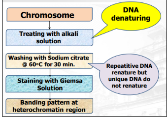
Color of Heterochromatic band in G-banding/Giemsa
Black
Heterochromatic band
condensation of heterochromatin
repeating sequences and has no role in transcription
does not become a protein because they are so tightly packed together that they do not enter the transcription factor
Color of Euchromatic band in G-banding/Giemsa
Lightly stained (white)
Euchromatic band
condensation of euchromatin
loose and can easily stick to transcription factor, that’s why it is easily converted into an mRNA
sequences that are present encode protein; protein coding genes
Color of Heterochromatic band in R-staining
Lightly stained (white)
Color of Euchromatic band in R-staining
Black
Reason why heterochromatin is tightly packed
Because it is methylated
Around how many base pairs wrapped around a histone
200 base pairs
Reason why euchromatin is loose
Because it is acetylated
Reagents for karyotyping
Glacial acetic acid
Methanol
KCl (hypotonic solution)
RPMI Growth Medium
Fetal Bovine Serum
Phytohemagglutinin
Colcemid
Giemsa Dye
Trypsin
Glacial acetic acid and Methanol
allow the cells to be fixed
Colcemid (Colchicin)
arrest to metaphase
Giemsa Dye
imparts color
KCl (hypotonic solution) (Potassium chloride)
cause the cells to become bigger/swell
RPMI Growth Medium and Fetal Bovine Serum
grow lymphocytes (White blood cells)
Phytohemagglutinin
destroys RBCs; stimulate mitosis
Trypsin
an enzyme that digest AT to make it stain (none of this will make it hard for Giemsa to stain)
Reason why we don’t need RBCs for karyotyping
Because it has no nucleus, thus no chromosomes
Part of cell where chromosomes are found
Nucleus
Materials needed for karyotyping
Sterile 5 mL syringe
21-gauge syringe needle
Conical tubes (15 mL)
Green-top Vacutube
Glass slides
Pasteur Pipette
Pipettor and Pipette tips
Serological pipettes
5 major steps in karyotyping
Short term lymphocyte culture
Harvesting
Fixing the cells
Making the chromosome slides
Slide Analysis
Use of antibiotics for karyotyping
To prevent microbial growth
Acronym for the 5 major steps in karyotyping (for Ero)
She Fucks MS
SHe Fucks MS
She Fucks MS abbreviation meaning
Short term lymphocyte culture
Harvesting lymphocytes
Fixing the cells
Making chromosome slides
Slide analysis
How long will the cultured blood cells have to be grown at an incubator at 37 degrees Celsius?
3 days
Temperature at which the cultured blood cells have to be in
37 degrees Celsius
After the addition of colcemid, how long will you incubate it?
15 minutes
Centrifuge settings when centrifuging in Harvesting lymphocyte stage
1000 RPM for 10 mins
Important portion in the tube for karyotyping
Pellet
What is contained in the pellet?
Lymphocytes
Where do we find the pellet in the tube for karyotyping?
Bottom
Reagent that is carcinogenic
Ethidium bromide, Actinomycin D, Bromodeoxyuridine (BrdU)
Ethidium bromide is the most carcinogenic
Cell synchronization
significantly increase the total yield of metaphase chromosomes. Cells are arrested at S phase by adding an excess amount of BrdU overnight (16 h). After this, the block is released by washing the cells and adding thymidine for 5.5 h before colcemid treatment
Centrifuge settings when centrifuging in the Fixing the cells stage
1200 RPM for 5 mins
In what major step do we use Carnoy’s fixative?
Fixing the cells
Carnoy’s fixative components
Absolute methanol: glacial acetic acid
3:1
Use of Carnoy’s fixative
So the cells will not undergo autolysis
(hindi masira)
How many times will Carnoy’s fixative step be repeated?
3 times
On the third time you repeat Carnoy’s fixative step, what do you do?
Incubate at 4 degrees Celsius for 10 mins
The most common method of staining chromosomes for differentiation which uses trypsin that digests the chromosomes at regions rich in basic amino acids (Arg and Lys).
GTG-banding (G-bands by Trypsin using Giemsa)
Where is trypsin from?
Extracted from pig’s large intestines
What basic amino acids does trypsin digest?
Arginine and Lysine
What nucleotides does trypsin digest?
Adenine and Thymine
Automated computer software to help view Karyogram
Cytovision by Applied Imaging Inc.
Why study binding patterns?
allow you to see smaller pieces of the chromosome, so that you could identify smaller structural chromosome abnormalities not visible on a routine analysis
Caspersson et al (1958)
Person who published their first paper describing the use of quinacrine mustard (fluorescent dye) to stain chromosomes thereby ushering in a new era of chromosome banding.
The Paris Report (1971)
The first attempt to provide nomenclature for chromosome banding in any species and thus its recommendations have been adopted to nonhuman species as well.
People responsible for Q (Quinarcine) banding technique
Casperson et.al
People responsible for G (Giemsa) banding technique
Summer et.al
People responsible for N (NOR) banding technique
Matsui & Sasaki
People responsible for C (Centromeric) banding technique
Line & Laursen
Q-banding use
stains AT-rich regions
AT>CG
G-banding use
Giemsa stain
AT-rich regions stained darker than GC-rich regions
Opposite of R-Banding
AT>CG
DARK; AT rich regions
LIGHT; GC rich regions
C-banding use
stains heterochromic regions close to the centromeres; usually stains the entire long arm of the Y chromosome
Heterochromatin is stained dark NEAR centromere
R-banding use
Opposite of G-Banding
GC-rich regions are stained darker than AT
GC>AT
Stain used in Q-banding
Quinacrine mustard
Advantages of Q-banding
simple and versatile
used where G band is not accepted
The most common is G banding but if it is not possible to use, you can use Q-banding.
used in study of chromosome heteromorphism
Disadvantages of Q-banding
tendency to fade during examination because it is dependent on fluorescence.
If the slides are not immediately examined, the fluorescence fades.
Photo-degradation
Chromophore - absorb light of a particular wavelength due to a chemical bond formed between dye and light
It can be non-specific
UV light breaks the chemical bond
TRUE OR FALSE: R-banding interacts with DNA with thiazine and eosin components of stain brightens sulfur rich regions
False;
Its G banding, not that technique
Advantages of G-banding
used in identification of bands rich in Sulfur content
used in the identification of chromosomal abnormalities
gene mapping
Disadvantages of G-banding
not used in plants
Methylene stains used for G-banding
Methylene Azure
Methylene Violet
Methylene Blue
Disadvantages of N-banding
Fumes caused by the acids
Trichloroacetic acid (TCA)
Hydrochloric Acid (HCl)
Advantages of N-banding
used in the identification of Nucleolar organizer region
superior banding pattern for plants
Identify the banding technique
C banding