


Introduction
Introduction
Old Phrenology
In the 19th century, Phrenology assigned separate functions to cortical areas
Bumps on the skull were thought to overlie enlarged brain regions which were matched to behaviors/skills/ personality
[Phrenology is the detailed study of the shape and size of the cranium (skull) based on the former belief that they are indicative of mental faculties and character]
Phrenologists are people who believed that the development of specific intellectual traits was associated with brain growth that literally made the skull overlying that part of the brain bulge out
By examining people believed to have high or low abilities in various traits and then associating these abilities (or lack thereof) with the relative height of various areas on their skull phrenologists created elaborate maps that identified the location of dozens of traits ranging from mathematical ability to caution
The fundamental premise of phrenology is now totally discredited
Certain areas of the brain do not map specifically or uniquely to certain types of intelligence
Nor do local areas of the brain (neocortex) grow and bulge out the skull according to the development of particular traits such as caution or secretiveness
The brain is not a diffuse non-differentiated association-forming blob
Clumps are called Nissl bodies
Stains are called Nissl stains
Origin of Modern Neuroscience
Neuron Doctrine
Neurons are specific cells that carry electrical impulses
The basic unit of the nervous system
Histology is the microscopic study of the structure of tissues
During the evolution of histology, the histologists could not properly distinguish between individual cells
German neurologist Franz Nissl found a way to selectively stain a cell’s nucleus and the surrounding area using cresyl violet
[Cresyl Violet: a basic dye and is used as a common stain in histology]
Nissl’s stains can help distinguish between neurons and glia as well as identify the arrangement of neurons
[Glial cells also known as glia is nonneuronal brain cells that provide structural nutritional and other types of support to the brain]
This arrangement of neurons in the brain is called cytoarchitecture
[Cytoarchitecture is the study of the structural arrangement of neurons within the central nervous system (neuronal size, shape, packing, density, and staining intensity are all features that are used to characterize a specific cytoarchitectural area region or trend]
Italian histologist Camillo Golgi used a silver chromate solution to create the Golgi Stain which focused on the neuronal cell body
{Note: Silver Chromate is an inorganic compound with the formula Ag2CrO4.}
[Golgi Stain is a tissue stain that completely flls a small proportion of neurons with a dark, silver-based precipitate.]
Golgi Stains helped determine the definitive characteristics of neurons including a central region which contains the nucleus and thin tubes that extend from the center
The central region is called the soma
These discoveries from Golgi Stains resulted in two opposing theories
Camillo Golgi
Golgi developed the reticular theory (which neurites of different cells form a continuous reticulum or network)
Spanish histologist Ramon y Cajal
Cajal argued that the neurites of different neurons are not continuous with each other and communicate by contact not continuity
Idea could be related to cell theory
[states that all living things are composed of one or more cells; the cell is the basic unit of life; and new cells arise from existing cells]
This concept that cell theory also applies to neurons is known as the neuron doctrine
[The neuro Doctrine is a concept that describes the properties of a neuron Is basically saying that the nervous system are separate discrete cells with processes arising out of the cell body]
Brain is composed of independent cells
Signals transmitted from cell to cell across gaps (Synapse)
[The junction between the presynaptic neurotransmitter squirting axon and the postsynaptic neurotransmitter-receiving dendrite]
The most fundamental computing element in the nervous system
Scientist believe that there may be up to 1015 (a quadrillion) synapses
The gap between the pre- and postsynaptic neurons is called the synaptic cleft
Golgi stains allow a view of only occasional cells
The cells that are selected are shown in their entirety including all of the dendrites and often the axon as well as the cell body
There are four tenets of the neuron doctrine
The neuron is the structural and functional unit of the nervous system
Neurons are individual cells;
Not continuous to other neurons neither anatomically or generically
The neuron has three parts: dendrite, soma (cell body) and axon
Soma (cell body) 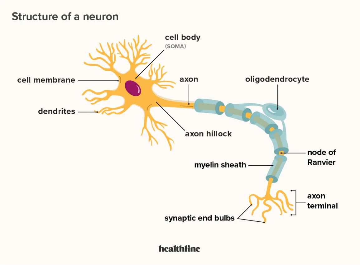
The thin tubes that radiate from the soma are called neurites
Can be either axons or dendrites
Somas usually have one axon
carrying the output of neurons
Numerous dendrites extend from the soma and receive the inputs of neurons
Neuron - anatomy
The Neuron consists of the dendrites, nucleus, axon hillock, cell body, myelin sheath, axon terminals, and the muscle fibers
Dendrites:
an extension of the cell body that receives information from other neurons (
Nucleus:
A collection of neuronal cell bodies within the central nervous system (e.g., the caudate nucleus)
[The caudate nucleus is a paired “C” shaped subcortical structure which lies deep inside the brain near the thalamus]
Axon hillock:
the cone shaped area in the cell body
Has unique properties that allows it to gather and integrate the information arriving from the synapses on the dendrites and cell body
Hollow tube
Includes various important substances such as enzymes and Structural proteins
These proteins are conveyed through the interior of the axon from the cell body (protein production) to the axon terminal (where they are used)
Cell body (soma):
the region of a neuron that is defined by the presence of the cell nucleus
Myelin Sheath:
A sheath that sounds the axons: provided by glial cells.
Axon Terminals:
Axon terminals is the button-like ending of an axon used by the axon, as a way to make synaptic contacts with other neurons or with the effector cell.
Muscle fibers:
A cylindrical cell composed of numerous myofibrils (rod like organelle of a muscle cell) that contracts when stimulated
Axon transport:
the transportation of materials between the cell body and the axon terminal
Works in both directions
Anterograde transport: moves materials towards the axon terminal
Retrograde transport: move material back to the cell body for recycling
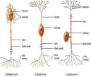
There are three kinds of neurons
Unipolar neurons
a single extension branches in two directions forming a receptive pole and an output zone
These are the simplest class of neurons
These neurons are usually sensory neurons with cell bodies located in the spinal and cranial nerve ganglia
Bipolar neurons
one axon one dendrite- usually sensory
These neurons usually runs from opposite sides of the cell body
Cells of this type are found primarily in the retina (eye) and elsewhere in the nervous system Can also be called a bipolar cell
Multipolar neurons
one axon many dendrites- most common type
This type of neuron is able to receive impulses from multiple neurons at the same time due to its many dendrites
The dendrites transmit the signals through the neuron via an electrical signal that is spread down the axon
The dendrites comprise the input zone
In multipolar and bipolar neurons the cell body also receives synaptic inputs (which makes it also a part of the input zone)
All neurons have four functional zones
Input
This is where neurons collects and integrate information either from the environment or from the other cells
Neurons receive information through synapses from other neurons
Some neurons have dendrites that are elaborately branched
Dendrites may be covered in dendritic spines
[small projections from the surface of the dendrite that add additional space for synapses]
Dendritic spines can change and increase surface area
Development of dendritic spine
Integration
This is where the decisions to produce a neural signal is made
This is where the neuron cell body (soma) combines (integrate-hence the name-) the receiving information to determine whether to send a signal or not
Conduction
This is where the information can be transmitted over great distance
This is where the axon (nerve fiber) carries the neuron electrical signal away from the cell body
Towards the end the axon may split into multiple branches (axon collaterals)
[axon collaterals: a branch of an axons]
Output:
This is where the neuron transfers information to other cells
Specialized swellings at the ends of the axon (axon terminal) transmit the neurons signals across synapse to other cells
There are two kinds of brain cells
Neurons
Basic unit of the nervous system
Each composed of receptive extensions (dendrites) integrating cell body a conducting axon and a transmitting axon terminal
Neurons are specialized cells that process information
Tiny dendritic spines have neural plasticity- their number and structure are rapidly altered by experience
[neural plasticity is the ability of the nervous system to change its activity in response to intrinsic or extrinsic stimuli by reorganizing its structure functions or connections]
Plasticity= brain cell can change
If the neurons was to break it cannot come back
If the brain was to be bigger it would not be able to work
Neurons use electricity to communicate what is happening in different parts of the neuron
Axons can conduct electricity
Brains are too fragile if a disease was to enter the brain it would damage their nerves
Glia
Glia- "glue" (Greek)
Glia are non-neuron cells (i.e not nerves) of the brain and nervous system
Provides physical and metabolic support to neurons including neuronal insulation and communication and nutrient and waste transport
Glial cells support the brain
More glial cells than neurons in the brain
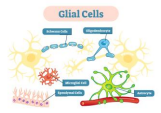
Four types of glial cells:
Oligodendrocytes and Schwann cells wrap around successive segments of axons to insulate them with fatty substance called myelin
[Myelin sheath is an insulating layer or sheath that forms around the nerves It has three functions: Its fatty-protein coating provides protective insulation for nerve cell It allows the electrical impulses to travel quickly and efficiently between one nerve cell and the next]
gives the myelin sheaths an axon the appearance of a string of elongated slender beads small uninsulated pages of axonal membrane nodes of Ranvier remain exposed Why does nodes of Ranvier remain exposed?
Astrocytes
"star"
Weave around and between neurons with tentacle- like extensions
Some stretch between neurons and fine blood vessels controlling local blood flow
Help to form the tougher outer membranes that swaddle the brain
Also secrete chemical signals that affect synaptic transmission and the formation of synapse
One of the three major classes of glial cells found in the central nervous system; important in regulating the ionic milieu of nerve cells and in some cases transmitter reuptake
What is astrocytoma?
Astroycytoma is a type of cancer that can form in the brain or spinal cord.
Alexander disease- astrocytes fill with gfap (in blue) the fail
Oligodendrocytes
Most numerous glial cell in the brain
Provide the blood-brain barriers
Regulate composition of the extracellular space
Wraps axons with myelin sheaths inside the brain and spinal cords (central nervous system)
Each oligodendrocyte wraps around several axons
[Nodes of Ranvier are gaps in the myelin sheath coating on the neural axon The nodes of Ranvier allows for ions to diffuse in and out of the neuron propagating the electrical signal down the axon]
After crossing the cleft the released neurotransmitter molecules interact with matching neurotransmitter receptors that stud the postsynaptic membrane
The receptors capture and react to molecules of the neurotransmitter altering the level of excitation of the postsynaptic neuron
affects the likelihood that the postsynaptic neuron will in turn release its own neurotransmitter from its axon terminals
Disease: Multiple sclerosis- Oligodendrocyte injury from autoimmune attack
Microglia
Extremely small motile glial cells that remove cellular debris for injured or dead cells
SER - smooth endoplasmic reticulum regulates cytoplasm
Ependymal Cells
Ependymal Cells Line ventricles secrete and absorb cerebrospinal fluid
Synapse
Information is transmitted from an axon terminal of a presynaptic neuron to the receptive surface to a postsynaptic neuron
Presynaptic Neuron- the transmitting side of synapse
Postsynaptic neuron- The region of a synapse that receives and responds to neurotransmitter
When the postsynaptic neuron is excited it fires off electrical pulses that travel away from its cell body and along its axon until they reach the places where the axon branches and the individual branches end (Axon terminals (also called terminal boutons))
Called spikes or action potentials
A Synapse can be divided into three principal components
The specialized presynaptic membrane of the axon terminal of the presynaptic (transmitting) neuron
[The presynaptic membrane is the specialized membrane on the axon terminal of a neuron that transmits information by releasing neurotransmitter]
The synaptic cleft ( a gap of 20-40 nanometer) that separated the presynaptic and postsynaptic neurons
[the space between the presynaptic and postsynaptic neurons at a synapse]
The specialized postsynaptic membrane on the dendrite or cell body of the postsynaptic (receiving) neuron
[The specialized membrane on the surface of a neuron that receives information by responding to neurotransmitter from a presynaptic neuron]
Presynaptic axon transmitter contain many tiny hollow spheres ( synaptic vesicles)
Each synaptic vesicle contains molecules of neurotransmitter the special chemical that presynaptic neurons use to communicate with postsynaptic cells
[Also called simply receptor A specialized protein that is embedded in the cell membrane allowing it to selectively sense and react to molecules of a corresponding neurotransmitter or drug]
Molecules of neurotransmitter generally do not enter the postsynaptic neuron
Simply bind to the outside of the receptors momentarily to induce a response and then detach and diffuse away
This communication start when in response to electrical activity in the axon synaptic vesicles fuse to the presynaptic membrane and then rupture, releasing their payload of neurotransmitter molecules in to synaptic cleft
Nervous System
Neuronal cell bodies, dendrites, axons and glial cell mass together to form the tissues that defines the anatomical features of the nervous system (gross neuroanatomy) that is visible to the naked eye
[Gross neuroanatomy is the anatomical features of the nervous system that are apparent to the naked eye]
Central nervous system is the brain and the spinal cord
The Peripheral nervous system is the portion of the nervous system that includes all nerves and neurons outside of the brain and spinal cord
[a collection of axons bundled together outside the central nervous system]
The Peripheral Nervous system (PNS)
There are three functional kinds of neurons
Sensory
Specialized to gather sensory information
Take on different shapes and sizes
This is the neurons that responds to environment such as light odor or touch
Motoneurons (motoneurons)
A neuron that transmits neural messages to muscles (or glands)
Large with long axons reaching out to synapses causing muscular contractions
Interneurons
This is the neurons that receive input from and send input to other neurons- integration (most neurons in CNS)
Most neurons in the brain are interneuron
Axons of interneurons only measure a few micrometers
Larger neurons:
Tend to have more-complex input and outputs
Cover greater distance
Convey information more rapidly than smaller neurons
Somatic nervous system
A part of the peripheral nervous system that supplies neural connections mostly to the skeletal muscles and sensory systems o f the body
Consists of cranial nerves and spinal nerves
Main pathway for the brain to control movement
12 pairs of cranial nerves
[a nerve that is connected directly to the brain]
Some of these nerves are exclusively sensory:
The olfactory (I) nerves transmit information about smell
The optic (II) nerves carry visual information from the eyes
The vestibulocochlear (VIII) nerves convey information about hearing and balance
Five pairs of cranial nerves are exclusively motor pathways from the brain:
The oculomotor (III) Trochlear (IV) Abducens (VI) nerves innervate muscles to move the eyes
The spinal accessory (XI) nerves control neck muscles
The hypoglossal (XII) nerves control the tongue
The remaining cranial nerves have both sensory and motor functions
The trigeminal (V) nerves for example transmit facial sensation through some axons but control the chewing muscles through other axons The facial (VII) nerves control facial muscles and receive some taste sensation
The glossopharyngeal (IX) nerves receive additional taste sensations and sensations from the throat and also control the muscles there
The vagus (X) nerve extends far from the head running to the heart liver and intestines and other organs
Its long convoluted route is the reason for its name
The vagus is the primary route by which the brain both controls and receives information from many visceral organs
Participates in such varied functions as sweating digestion and heart
31 Pairs of spinal nerves
[also called somatic nerve: a nerve that emerges from the spinal cord]
Emerges through regularly spaced opening along both sides of the backbone
Each nerve is made up of motor fibers- projecting from the ventral (front) of the spinal cord to the organs and muscles
Named according to the segments of the spinal cord 8 cervical (neck)
12 thoracic (torso)
5 lumbar ( lower back)
5 sacral (pelvic)
1 coccygeal (bottom)
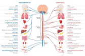
Autonomic nervous systems
A part of the peripheral nervous system that provides the main neural connections to the internal organs.
Has two divisions
Sympathetic
Parasympathetic
act in opposite fashion.Axons of the sympathetic nervous system exit from the middle parts of the spinal cord and innervate he sympathetic ganglia (small cluster of neuron for inside the CNS)
Sympathetic innervation prepared the body for immediate action (fight or flight)
Blood pressure increase
The pupils of the eyes widen
The heart quickens
Norepinephrine comes from sympathetic nerves
Acetylcholine from parasympathetic
The balance between the two systems determines the state of the internal organs
Parasympathetic activation rests and digest
Helps body to relax, recuperate and prepare for future action
Nerves of the parasympathetic system originate in the brainstem and in the sacral spinal cord
Preganglionic neurons (blue) from CNS to autonomic ganglia
Postganglionic neurons (red) from autonomic ganglia to target in the body
Anatomical convention for the anatomy of the brain
To describe the flow of neural information:
Afferent – carries impulses into a region of interest (sensory)
Efferent – carries impulses away from a region of interest (motor)
Coronal:
separate the brain from front to back(resembles a butterfly) (crown
Sagittal (midsagittal):
slices the brain down the midline so you can see what's on each half (side view)
Horizontal:
separates the brain from top to bottom
White vs gray matter
White matter is composed of axon bundle (the neural “wires” that connect neurons to each other)
It is white because the myelin sheaths (which are white fatty tissues) cover the axons
Axons live in the white matter
White matter tracts (Axons) connects brain areas
Several long tracts run in an anterior- posterior direction
Short tracts arch between nearby areas of the cortex
Long projection fibers run to and from the cerebral cortex
Some go through the corpus callosum connecting homologous regions of the two hemispheres
Grey matter is composed of cluster of neuron cells bodies
Is gray because it has a dark gray appearance
Cell body lives in the gray matter
(neuronal cell bodies and dendrites which receive inputs from other neurons and do the neural computations)
Cell bodies= inside
Axons= outside
The brain has more white matter than gray matter
Why? The brain uses local interconnections between neurons to do computations in neural circuits