


1.2: Cell structure and organisation
Cell structure
The detailed structure of a cell is known as the ultrastructure, and is only visible through a microscope.
There are multicellular organisms - contain multiple cells, unicellular - contain one cell and acellular - contain no cells.
There are two types of cells, prokaryotes which are simpler cells without a nucleus and eukaryotes which have membrane bound organelles and a nucleus.
All cells have a thin membrane made of proteins and phospholipids. Eukaryotes have membranes around their organelles for a larger surface area due to their large surface area and ability to provide a transport system.
Plant and animal organelles
Nucleus
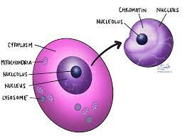
This contains the DNA which comprises the chromosomes with protein. They are the site of transcription, and provide a template for DNA replication.
It has many parts:
Nuclear envelopes - these are a double membrane which surround the organelle, with pores to allow large molecules such as mRNA and ribosomes out. The outer membrane is linked to the endoplasmic reticulum (ER).
Nucleoplasm - this is the granular material inside the nucleus containing chromatin, which is made of coils of DNA bound to proteins. This condenses to chromosomes during cell division.
Nucleolus - these are one or more spherical bodies inside the nucleus that synthesises rRNA, a component of ribosomes.
Mitochondria
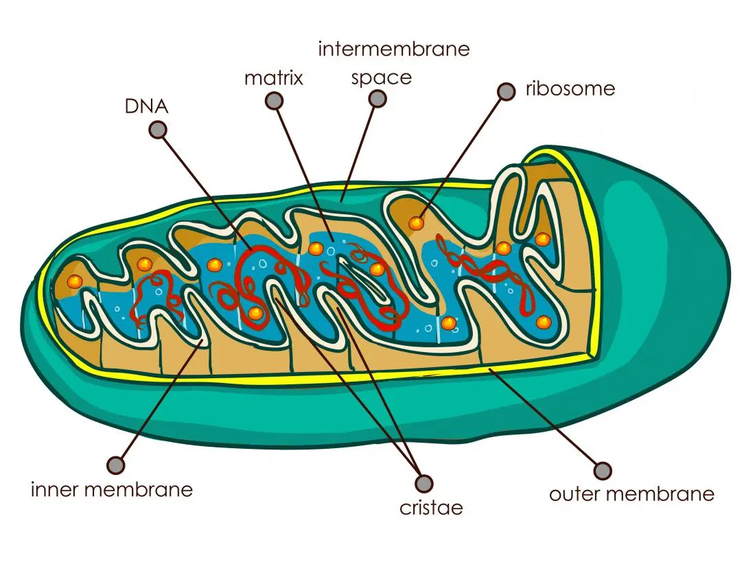
These produce ATP in aerobic respiration, with reactions occurring across the organelle. They are cylindrical to increase surface area.
There are two membranes separated by a intermembrane space, the inner one folded inwards to form cristae.
An organic matrix, which is a solution containing many compounds, including lipids and proteins.
Small circles of DNA, so they can replicate and code for some proteins and RNA.
Small 70S ribosomes to allow for protein synthesis.
Their function is to produce ATP in aerobic respiration. These reactions can occur in the matrix or on the inner membrane.
Endoplasmic reticulum
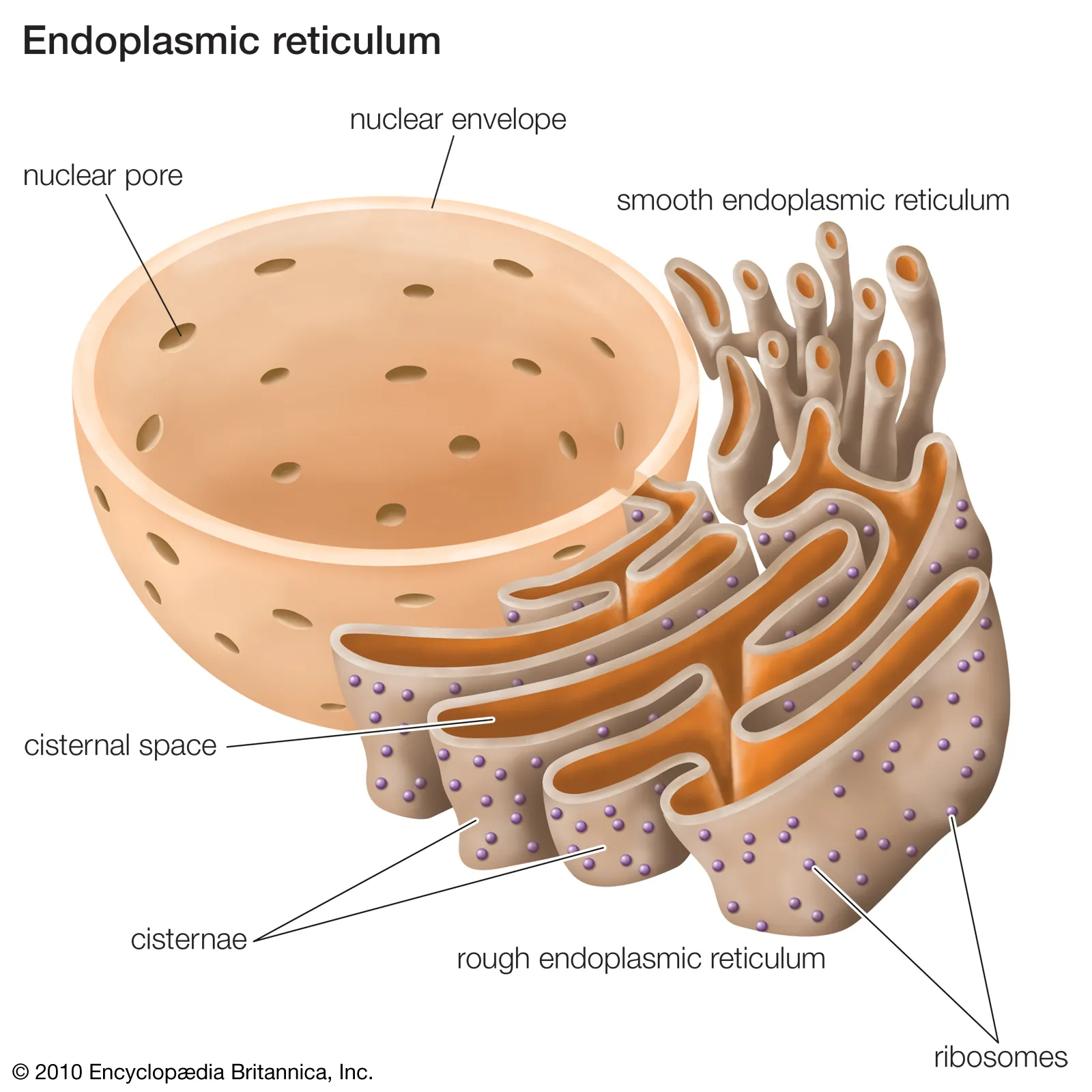
ER is a system a parallel double membranes forming flattened sacs with interconnected, fluid-filled spaces known as cisternae. It is connected to the nuclear envelope.
It allows for the transport of material through the cell, and there are two types:
Rough ER (RER) has ribosomes on the outer surface and transports the proteins made by them.
Smooth ER (SER) has no ribosomes. It is associated with the synthesis and transport of lipids.
Ribosomes
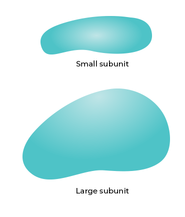
Ribosomes have two subunits, one large subunit with 2 tRNA attachment sites and one small subunit with one mRNA attachment.
They are assembled from ribosomal RNA (rRNA) and protein. They are the site of translation, where mRNA and tRNA are used to assemble the polypeptide chain.
They are smaller in prokaryotic cells than eukaryotic cells, 70s vs 80s.
Golgi body
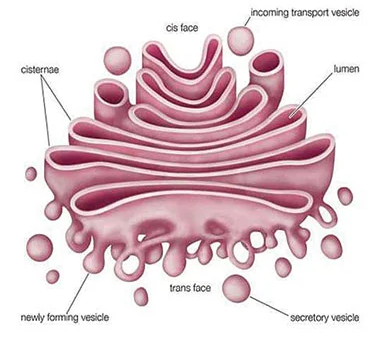
It resembles a more compact version of the ER.
Vesicles containing polypeptides pinch off from the RER and fuse with the golgi body.
They are modified and packaged by the golgi body, before being pinched off at the other end of the golgi body.
These vesicles may carry proteins elsewhere in the cell or secrete through the cell membrane via exocytosis.
Their functions include:
Producing secretory enzymes.
Secreting carbohydrates.
Producing glycoprotein.
Transporting and storing lipids.
Forming lysosomes.
Animal only
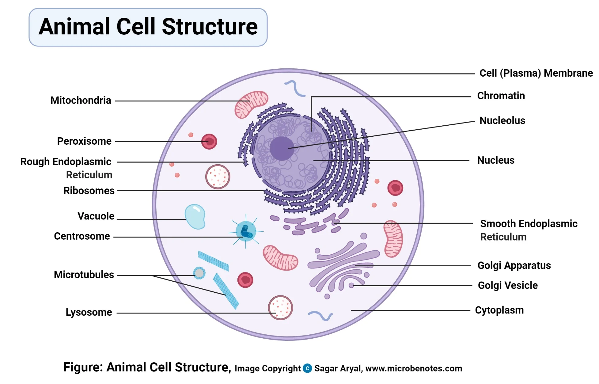
Lysosomes
These are small, temporary vacuoles surrounded by a membrane.
They are formed from being pinched off a golgi body.
Their role is to contain and isolate potentially harmful digestive enzymes from the rest of the cell, and only release them to recycle worn out organelles.
They can also digest material that has been taken into the cell, such as when a white blood cell engulfs bacteria by phagocytosis.
Centrioles
They are located just outside the nucleus, and together are known as the centrosome.
They are two rings of microtubules, and make hollow cylinders positioned at right angles to one another.
During cell division, centrioles organise the microtubules that make the spindle.
Plant cell only
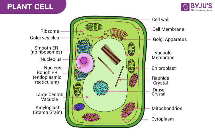
Chloroplasts
These occur in the cells of photosynthesising tissue, with the highest concentration closest to the sun.
Each chloroplast is surrounded by two membranes, called the chloroplast envelope.
The stroma is fluid filled, containing some products of photosynthesis such as lipid droplets and starch grains. This can take up a large part of stroma.
Chloroplasts contain 70S ribosomes and circular DNA which enable them to self-replicate and make their own proteins.
Vacuole
A large permanent fluid-filled sac bounded by a single membrane known as the tonoplast.
They contain cell sap, which stores chemicals such as glucose, amino acids, minerals, and may store vitamins and pigments.
They have a major role in supporting soft plant tissues.
Cell wall
It consists largely of cellulose, which are held in microfibrils, which are aggregated into fibres, embedded in a polysaccharide matrix called pectin. They have many functions, such as:
Transport - there are gaps between the cellulose molecules which make the cell wall fully permeable to water, dissolved molecules and ions.
Strength - the structure of the cellulose microfibrils supports the cell structure, especially when the cell is turgid.
Communication between cells - plasmodesmata, which is strands of cytoplasm which can only occur where the is no cellulose thickening between two cells. The run between cells to allow water transport. The network of plasmodesmata is known as the symplast.
Endosymbiotic theory
This is a theory on the origin of chloroplasts and mitochondria, that says at least 1.5 x 10*9 years ago bacteria with fluid membranes absorbed other bacteria and formed a symbiotic relationship.
This is evidenced by chloroplast division closely resembling that of free-living cyanobacteria.
Prokaryotes, eukaryotes and viruses
Introduction
Prokaryotes are the least advanced cells, and therefore resemble the first living cells most closely. Examples are bacteria, algae and Archaea.
Eukaryotes evolved from prokaryotes, and have the most advanced structure. Examples are animals, plants and fungi.
Viruses are not made of cells, and therefore not classified with living organisms.
Prokaryotes
Prokaryotes have no nucleus or internal membranes, meaning there are no membrane bound organelles such as the mitochondria, chloroplast or vacuole.
They rarely form multicellular structures, and are often known as unicellular.
Prokaryotes have a few different parts:
Their DNA is loose in the cytoplasm, and there are small plasmids throughout containing DNA.
They have a peptidoglycan cell wall.
They still have a cell membrane, 70s ribosomes, cytoplasm.
They are also smaller than eukaryotes.
Only some prokaryote contain:
Mesosomes can perform aerobic respiration.
Photosynthetic lamellae hold photosynthetic pigments, and can carry out photosynthesis or nitrogen fixation.
Flagellum which allow the cell to move around.
Pili, which allows them to attach to other cells during sexual reproduction.
Capsule, or slime coat, is a sticky surface that protects the cell.
Viruses
They are described as acellular, as they are not made of cells. They have no organelles, DNA or cytoplasm.
When they are outside of cells they are known as inert virions, and once infected a host cell they take over its metabolism and multiply inside it.
They have a core of nucleic acid, either DNA or RNA, surrounded by a protein coat known as a capsid. Sometimes a membrane derived from the host cell surrounds the capsid.
All cells can be infected with viruses, and ones that attack bacteria are known as bacteriophages.
Viruses can be crystallised, and their only trait of a living organism is the ability to reproduce.
Examples of diseases are HIV, flu, avian flu, swine flu and chickenpox.
Levels of organisation
Multicellular organisms have specialised cells, which form different structures and roles. The process of them becoming specialised is known as differentiation. During this process, cells change in structure and in the chemical reactions they perform.
Stem cells, however, have the potential to become any cell type in the body.
These specialised cells work together to form structures, which are listed below:
Tissues
Epithelial tissue
This tissue forms a continuous layer, covering or living internal or external surfaces of the body.
They can have nerve endings but no blood vessels.
Epithelial tissue cells sit on basement membranes, made of collagen and protein and vary in shape and complexity.
They usually have protective or secretory functions.
The types of epithelial tissue are:
Cuboidal epithelium - the simplest type, where they form cuboidal shapes and are only one cell thick. They occur in the ducts of salivary glands.
Columnar epithelium - has elongated cells, taller than they are wide, with cilla to increase surface area. They occur in the small intestine.
Squamous epithelium - flattened, thin cells. Forms walls of the alveoli.
Stratified epithelium - made from multiple layers, with the top ones dead to avoid damaging the ones underneath. Forms the skin.
Ciliated epithelium - moves substances and particles using cilla, hairlike projections that move in a wavelike manner. For example in fallopian tubes.
Muscle tissue
There are three main types of muscular tissue:
Skeletal tissue - this is attached to bones and generates locomotion in mammals. It has bands of fibres which gives powerful contractions. However, the muscle can tire easily. They are known as voluntary muscles, as they are controlled by the organism. They are also known as striated, as you can see stripes on them.
Smooth muscle - has individual spindle-shaped cells that contract rhythmically, and doesn’t tire easily. However, they contract less powerfully. They occur in the skin, such as in digestive tracts. They are known as involuntary, as they are not controlled by the organism. They are also known as unstraited, as they do not have stripes.
Cardiac muscle - only found in the heart. It is striated, involuntary, never tire and lack fibres such as in skeletal tissue. Their movement requires no nerve or hormone stimulation, however it can be altered by these factors.
Connective tissue
Connects, supports or separates tissues and organs. It is made up from elastic and collagen fibres in an extracellular fluid or matrix. Between cells are adipose, which is fat-storing cells, and cells of the immune system. Examples are:
Fibrous connective - found in tendons and joins muscle to bone.
Loose connective - found between layers of tissue.
Organs
These are made out of several tissues working together to perform a specific function. Examples are eyes in humans and leaves in plants.
Organ systems
A group of organs working together with a particular role, such as the reproductive system in both plants and animals.
Organisms
All the organ systems in the body working together to make an organism, such as a human or tree.