
R Chapter 2 - Cell injury, Adaptation and Death
History
Hippocrates( 460-337 BC): diseases result from form disturbed balance between he found body fluids
blood
phlegma(thick mucus)
black bile
yellow bile
Father of pathology → Rudolf Virchow
know for the idea that diseases stem from changes in healthy cells and that each disease only affects a certain set of cells rather than the entire organism
Terms
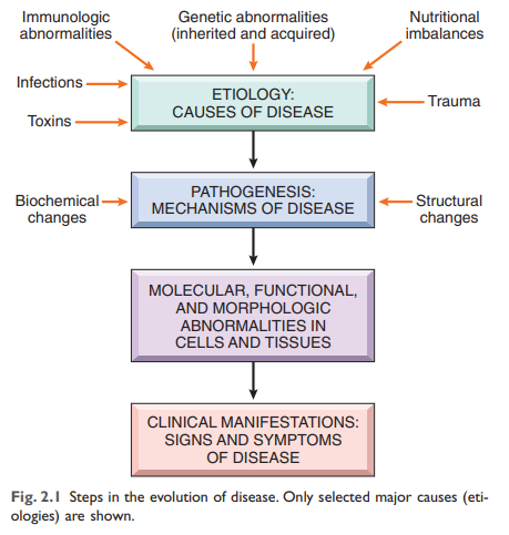
Pathology: the study of diseases → investing the causes of diseases, the associated changes at the levels of cells, tissues
Etiology: Origin of the disease, including underlying causes and modifying factors
Pathogenesis: the mechanisms of development and progression of disease
describes how etiological factors trigger molecular and cellular change giving rise to specific structural….
Causes of Cell Injury
Hypoxia: oxygen deficiency
most common cause: arterial obstruction
Ischemia: reduced blood supply (not only liquid loss but also essential nutrients loss and build-up of waste products)
Toxins
air pollution
insecticides
CO
Asbestos
cigarette smoke
ethanol
Drugs(medical and street drugs)
Infectious agents
virus
bacteria
fungi
protozoans
Immunologic reactions
autoimmune (reactions against one’s own tissues)
allergies (reactions against environmental substances)
inflammatory reaction
Genetic abnormalities
deficiency in proteins
protein abnormalities
triggered cell death
Nutritional imbalance
physical agents
Trauma (accidents)
extreme of temperature
radiation
electric shock
changes in atmospheric pressure
Aging
during cells life, their ability to respond to stress decreases causing irreversible damaging → leading to death
Stress
Sequence of vents in cell injury and cell death
Cellular reversible injury
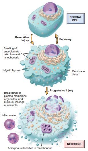
Reversible injury: is the state of cell injury at which the deranged function and morphology of the injured cells can return to normal if the damaging stimulus is removed
the cell grows in size( cellular swelling) to maintain an equilibrium since energy depend on ion pumps start failing, causing a disbalance in the cytoplasm
Fatty changes are the manifestation of lipid vacuoles
cell becomes more “purple.”
blebbing
three phenomena → Point of no return to cell death
inability to restore mitochondrial function
loss of structure and functions of the plasma membrane and intracellular membranes
loss of DNA and chromatin structural integrity
Cell Death
Causes for cell death
loss of oxygen and nutrient supply → called ischemia
actions of toxins
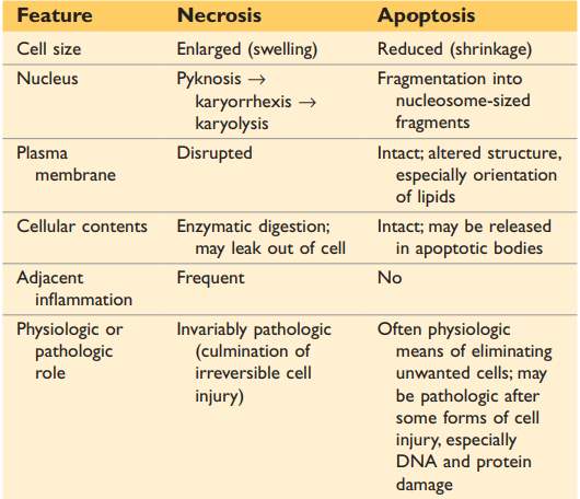
Necrosis
Necrosis: uncontrollable form of death
resulted from severe damage and caused the membrane to burst, and intracellular enzymes treat leaking out, causing the cell to digest itself
does not occur in healthy tissue; only occurs in pathologic processes
Types of tissue necrosis
Coagulative necrosis → blockage of blood -→ tissue dies
Liquefactive necrosis → necros in the brain often causes liquefactive
gangrenous necrosis → infection of bacterium that causes fast necrosis + swelling of legs + low temperature
Caseous necrosis → when a secluded area, that experiences necrosis the macrophages are locked in causing the tissue to look like cjees
Fat necrosis → when fat suddenly dies because when you have pancreatitis the enzyme that break down fat, start digestion fat storages → causing fat break down all over your bod<
Fibrinoid necrosis
Apoptosis
Apoptosis: controlled cell death
clears cell fragments in vesicles to prevent inflammatory reactions that could damage neighboring cells triggered by the enzymes called caspases
causes of apoptosis
Physiological triggers that happen in healthy bodies
loss of growth factor signaling
cells become too old
Pathologic
DNA damage
accumulation of misfolded proteins
Infections
pathways of Apoptis
intrinsic → mitochondrial pathway triggered by growth factor withdrawal, DNA damage or protein misfolding
all elements for apoptosis are found in mitochondria which develops the apoptosis,e. The apoptsome activates with Caspase 9 which causes cell death
Extrinsic → death receptors pathway
important receptor-ligand interaction: Fas and TNF receptors
when cells are old or broken, they start developing receptors called Fas. When fas ligand bond to the death receptors, apoptosis begins
caspapes 8 is also involved
Autophagic vacuoles consume cells
phosophatidlysis break down the cells in an controlled manner
Clearance of apoptotic cells
tidylserine on the inner side of the cell membrane triggers a “eat me” signal. In apoptotic cells, the membrane is flipped and tissue macrophages recognize the cell
apoptotic cells also secrete soluble factors that recruit phagocytes
SLIDE graph!!
Other Pathways of Cell Death
Necroptosis: TNF receptors and receptor-interacting protein( RIP) kinases are activated, initiating a series of events that result in the dissolution of the cell, much like necrosis.
Pyroptosis: a highly inflammatory form of lytic programmed cell death that occurs most frequently upon infection with intracellular pathogens and is likely to form part of the antimicrobial response (wiki def. )
Autophagy
Autophagy: (“self-eating”) refers to lysosomal digestion of the cell’s own compartments
survival mechanism during nutrient deprivation
Mechanisms of cell injury and cell death
The cellular response to injurious stimuli depends on the type of injury, its duration, and its severity
The consequences of an injurious stimulus also depend on the type, status, adaptability, and genetic makeup of the injured cell.
Cellular adaptations to stress
Adaptations are reversible changes in the number; size, phenotype; metabolic activity; o functions of cells n response to changes in their environment
cell under stress
when cell is under stress, the proteins are more misfolded, the ER has an adaptive unfolded response and in the worst case terminal unfolded response ( each tissue has different amount of senores (terminal UPR))
the amino acid sequence is as important as the the 3D shape of the protein
Shape dirctes function!!
CHaporone Sytsem→ system in ER that help fold proetins correctly in times of stress
chaperones interact with fromin proteins that hold the protein place to stabilize
some chaperones hydrolyze(split) the protein to proofread the amino acids ( histok proteins)
termalize out side of the cell in protoomes
Sensores
sensors that trigger increased folding
senores that trigger the stoping of producing proteins
senores that increase the chaperoning system (paradox)
some protins are not stoped because they have to continue caporoing
Adaptation Causes
physiological adaptations: responses of cells to normal stimulation by hormones or endogenous chemical mediators
for example, hormone-induced enlargement of the breast
pathologic adaptation: response to stress that allows cells to modulate their structure and function and thus escape injury
How can the cell change
Hypertrophy: increase in the size of cells resulting in an increase in size of organ
result of either physiological or pathological stimuli and is cause by increased functional demand or by growth factor or hormonal stimulation( growth factors)
Hyperplasia: increase in cell number due to increased proliferation
hormonal hyperplasia ( example growing of breasts during puberty due to hormonal changes)
compensatory hyperplasia residual tissue grows after removal or loss of an organ
Atrophy: shrinkage in the size of cells by the loss of cell substance
causes: decreased workload; diminished blood supply ; inadequate nutrition ; loss of endocrine stimulation ; aging
Cellular atrophy results from a combination of decreased protein synthesis and increased protein degradation.
Metaplasia: change in which one adult cell type (epithelial or mesenchymal) is replaced by another adult cell type( not pre-cancerous)
stemcell makes new cell that can better withstand the stress
Dysplasia: cells have DNA defects which can cause to cancer (pre-cancerous-)
SLide : metaplasia vs Dsplasia
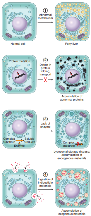
Intracellular accumulation
Under some circumstances, cells may accumulate abnormal amounts of various substances, which may be harmless or may cause varying degrees of injury. The substance may be located in the cytoplasm, within organelles (typically lysosomes), or in the nucleus, and it may be synthesized by the affected cells or it may be produced elsewhere.
Fatty Change: accumulation of triglycerides within the cells
Cholesterol and Cholesteryl Esters: phagocytic cells may become overloaded with lipid
Proteins: accumulation of abnormal proteins in cell
glycogen: Excessive intracellular deposits of glycogen
Pathologic calcifications
Dystrophic calcification: deposition of calcium at sites of cell injury and necrosis
Metastatic calcification: deposition of calcium in normal tissues caused by hypercalcemia (usually a consequence of parathyroid hormone excess)
Cellular Aging
cause the result of progressive decline in the lifespan and functional activity
abnormalities contribute the aging of cells
Accumulation of mutations in DNA
Decreased cellular replication
Defective protein homeostasis
Persistent inflammation
@path_logos
cellular response to stress
physiological adaptation
hyperthropia increase in cell volume
hyperplasia increase in number of cells
Class notes
shut down the process when 02
→ slide
Roles of Aptosis
death is as important as proliferation
for example: removal of tissues (tail that degenerates in frogs life span) + organ sculpting
apoptosis is a key role in regeneration
wind factors are excreted when a cell is destroyed and promotes new tissue to produce
if there is necrosis the tissue can be replaced only with scare tissue that does not have the function of previous tissue
apoptosis and the immune system
1 T lymphocyte recognizes one foreign antigen
look at thymic selction/deletion
when cell is infected by a virus( or something els) t cells can trigger apoptios in the cell to control the development of the disease
tuberculosis can survive weeks in macrophages( because it is resistant to acids)
deasis that are trigger defected apoptosis -→ look at picture in
SLIDE
chronic inflammation
cancer
auto immune disease
Infalamtory
IBD
viral infecation
Adeo virus
other factors that cause apotois
starvation
Lack of growth factor fo neurons
CD-25 receptor
LAck of negative feedback by endocrine system
developmental involution
a strutere was there before and grew together again
Necrosis coagulation
blood build up and the body preserves
Diabetes
pre dieabetis: when serving cell still produce enough insulin for the body due to increase production from the healthy pancreatic cells (called honeymoon phase). After weeks/years of overproduction the helathy cells die or make mistakes because of the long stress.