
BIO 1ST QT
HOMEOSTASIS AND FEEDBACK MECHANISM
Homeostasis
the regulation and maintenance of the interval environment of the body
conditions within the body must remain within a narrow range – like your body temperature
a. homeostasis involves keeping the internal environment within set ranges
b. control system help maintain homeostasis
- sensors gather data
- control centers receives data and sends messages
- communication system delivers messages to target organs, tissues
- targets respond to change
Feedback mechanism
Components of feedback loops:
- stimulus – it is something in the environment that causes a change
- receptor – it is a cell or tissue that receives stimulus
- relay- it is a transmitter of the message via nerves or hormones or both to the effector
feedback loops
feedback compares current conditions to set ranges.
a. negative feedback loops are necessary for homeostasis and stops the change
b. positive feedback loops promote growth hormones and stimulate cell division it also increases change
RESPIRATORY SYSTEM
A. FUNCTION: Exchange of gases
B. PHASE OF RESPIRATORY:
1. Exchange of gases between blood and air (external
respiratory)
2. Exchange of gases between blood and cells (internal respiratory)
C. PARTS:
1. Nose
- Internal part in the skull above the roof of the oral cavity; external part protruding from the face
-Opens to the exterior through the anterior nares and to the nasopharyngeal posterior nares
-Floor formed by the palatine bones and maxillae
-Lined with ciliated mucosa
-Serves as the passageway for incoming and outgoing air filtering, warming, and moistening it -Sense organ of smell
2. Pharynx Divided into:
a. Nasopharynx – behind the nose with four opening, two auditory or Eustachian and two posterior nares - Tonsils found in the nasopharynx destroys incoming bacteria and detoxify foreign protein
b. Oropharynx – behind the mouth with one opening, filtering from the oral cavity
c. Laryngopharynx – behind the pharynx with two openings, into the larynx and into the esophagus
- Serves as passageway into respiratory and digestive tracts
3. Larynx
- Box-like cartilaginous structure located just below the pharynx
Consists of:
a. Thyroid cartilage or “Adam’s apple" - protects the vocal chords behind it
b. Epiglottis or “lid cartilage” - a flap in the throat that keeps food from entering the windpipe and the lungs
c. Cricoid or “signet ring” cartilage -functions as an attachment site for muscles, cartilage, and ligaments involved in opening and closing the airway and in producing speech
d. Arytenoid or “pyramid shaped” cartilage -attachment to vocal cords that allows and aid the vocal cords’ movement
Vocal cords of two types:
1. False vocal cord (vestibular folds)
- folds of mucus lining
2. True vocal cord – fibro
- Elastic bands stretched across the interior of the larynx
- Slit between vocal cords in the glottis through which air enters and leaves the lower respiratory passages.
4. Trachea
- -Cartilaginous tube 10
- 11 cm. in length extending from the larynx to bronchi
- -Lined with ciliated mucosa
- For passageway of air, to and from the lungs
5. Lungs
- -Cone-shape organs that completely fill the pleural spaces extending from slightly above the clavicle to the diaphragm where the base of the lungs rest
- Covered by visceral pleura
Structures associated with lungs:
a. Bronchi–right and left formed by branching of trachea
b. Bronchioles – smaller branches of bronchi
c. Alveolar ducts–microscopic branches of bronchioles
d. Alveoli – microscopic sacs at the ends of the alveolar ducts provided with a network of lung capillaries
- Serves for the exchange of gases between blood and air.
D. Amount of Air Exchanged in Breathing - Measured by apparatus called spirometer
a. Tidal Air - average amount expired after a normal respiration of 500ml.
b. Expiratory reserved volume (ERV) – the largest additional volume of air that can be forcefully expired after a normal inspiration and expiration; 1,000- 1,200ml.
c. Inspiratory reserved volume (IRV) – largest addition of a volume of air that can be forcibly inspired after normal respiration; 3,000 – 3,300ml.
d. Residual Air – that which cannot be forcibly expired from lungs; about 1,200 ml.
e. Minimal Air – that which can never be removed form alveoli if they have been inflated even once, even though lungs are subjected to atmospheric pressure that squeezes part of the residual air out.
f. Vital Capacity – approximate capacity of lungs as measured by the amount of air that can be forcibly expired after forcible inspiration; varies with the size of the thoracic cavity, which is determined by various factors (size of rib cage, size of the heart).
E. Types of Breathing:
1. Eupnea/Eupnoea – normal quite breathing
2. Apnea – temporary cessation of breathing
3. Dyspnea – difficult breathing
4. Orthopnea–inability to breathe easily in a horizontal position
5. Tachypnea–excessively rapid and shallow breathing
6. Bradypnea- abnormal slow breathing
MUSCULAR SYSTEM
A. FUNCTIONS:
- Movement
- Posture
- Heat production
B. PROPERTIES OF MUSCLES
- Excitability
- Contractibility
- Extensibility
- Elasticity
C. TYPES OF MUSCLES:
1. Striated Voluntary or Skeletal Muscle
- Attached to the skeleton
- Controlled by the voluntary nervous system via somatic motor nerves in the spinal and some cranial nerves
2. Smooth Involuntary or Visceral Muscle
- Located in muscle layer of visceral organs
- Controlled by autonomic nervous system via autonomic motor neuron in autonomic, spinal, and some cranial nerves
3. Striated Involuntary or Cardiac Muscle
- Found in the heart
- Control is identical to that of smooth muscle
D. THE SKELETAL MUSCLE
- Typically spindle–shaped and composed of long muscle cells referred to as muscle fibers bound together by a connective tissue called fascia
- Each muscle contains several muscle bundles or fasciculi
E. MUSCLE FIBER CONTRACTION
Muscle - Muscle Fiber - Sarcomere
Steps:
a. PERIOD OF STIMULATION - Electrical energy transmitted along transverse tubules
b. PERIOD OF CONTRACTION - Calcium ion released inactivates troponin which normally blocks interaction between myosin and actin. ATP released causes actin to slide along myosin filaments.
c. PERIOD OF RELAXATION - Calcium ions are pumped out of the sarcoplasm back into the sarcoplasmic reticulum, returning the muscle fiber to their resting state
- When sarcomeres are fully contracted, the appearance of thick and thin filament changes.
F. Type of muscles as to Action:
Flexor–bends a part towards another
- Ex. Biceps brachii
Extensor – straightens a part
- Ex. Triceps brachii
Adductor–draws a part towards the median line
- Ex. Pectoralis major, Adductor longus
Abductor – draws a part away from the median line
- Ex. Deltoid, Supraspinatus
Levator–raise a part
- Ex. Levator Scapulae
Depressor–lowers a part
- Ex. Trapezius, Serratus anterior
Constrictor – closes an opening
- Ex. Sphincter pupillae
Dilator – works against a constrictor
- Ex. Dilator pupillae
Pronator – bends back of hand forward
- Ex. Pronator teres, Pronator quadratus
Supinator – bends the palm forward
- Ex. Supinator muscle
Rotator – turning a body part around its own axis Circumduction - Moving in a circle at a joint
G. METHODS OF ATTACHEMENT TO BONES:
1. Tendon- Strong, tough connective tissue cord
2. Fascia- Tough, sheet like Membrane which covers and protects tissue
H. ORIGIN AND INSERTION:
Origin: end that does not move
Insertion: end that Moves when muscle contracts
I. TERMS ASSOCIATED WITH MUSCLE AND MUSCLE CONTRACTION
1. Hypertrophy – physical enlargement of a muscle due to the addition of more myofibrils to the muscle fibers making them swell.
2. Atrophy – reduction in the size of a muscle due to the decrease in the number of myofibrils in the muscle fibers.
3. Treppe (Staircase Phenomenon) – when a muscle has contracted a few times, subsequent contractions are more powerful
4. Shivering – rapid, repetitive, involuntary skeletal muscle contractions stimulated by a hypothalamic temperature-regulating center.
5. Cramps – caused by sustained muscle contraction. 6. Rigor Mortis – strong association of actin and myosin after death due to ATP depletion.
J. BURSAE
- Small sacs lined with sinusoid membrane and contains synovial fluid.
- Located whenever pressure is exerted over moving parts like between skin and bone, tendons and bone, muscles or ligaments and bones.
- Act as cushions relieving pressure between moving parts
Tissues
Introduction:
- Cells are arranged in tissues that provide specific functions for the body.
- Cells of different tissues are structured differently, which leads to their differences in function.
- The tissues of the human body include four major types
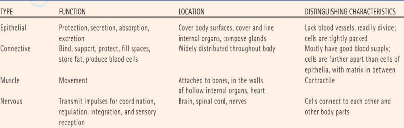 These four tissue types have a wide range of functions, as shown in the following table.
Epithelial Tissues:
A. General Characteristics
1. Epithelial tissue is widespread throughout the body, covers organs, and lines body surfaces.
FUNCTIONS
- Protection
- Absorption
- Sensation
- Reproduction
Types of Epithelial Tissues
Simple Squamous Epithelium - is made up of a single layer of thin, flattened cells.
Simple cuboidal epithelium - consists of a single layer of cube-shaped cells with centrally located nuclei.
Simple columnar epithelium - is made up of a row of elongated cells whose nuclei are all located near the basement membrane. It may be ciliated.
- In the intestine, these cells possess microvilli that increase the surface area available for absorption
- Mucus-secreting goblet cells can be found among columnar cells.
Pseudostratified Columnar Epithelium - these cells appear layered due to the varying positions of their nuclei within the row of cells, but are not truly layered.
Stratified Squamous Epithelium - this type of tissue is made up of layers of flattened cells that are designed to protect underlying layers
- In the skin, outer layers of cells undergo keratinization; however, this process does not occur where tissues remain moist in the throat, vagina, or anal canal.
Stratified Cuboidal Epithelium - this tissue consists of two to three layers of cuboidal cells lining the lumen of the mammary glands, sweat glands, salivary glands, and pancreas.
Stratified Columnar Epithelium - this tissue consists of several layers of cells and is found in the vas deferens, part of the male urethra, and parts of the pharynx.
Transitional epithelium - is designed to distend and return to its normal size, as it does in the lining of the urinary bladder.
Glandular Epithelium - this tissue is made up of cells designed to produce and secrete substances into ducts or into body fluids.
- Glands that secrete products into ducts are exocrine; those that secrete into body fluids and blood are called endocrine.
Glands are classified by the ways they secrete their products:
a. Merocrine glands release fluid products exocytosis (pancreas) and are grouped as serous which produce a watery fluid or mucus which produce a thicker, protective substance.
b. Apocrine glands lose portions of their cell bodies during secretion (sweat glands).
c. Holocrine glands release entire cells (sebaceous glands).
Connective Tissues:
A. General Characteristics
1. Connective tissues bind, support, protect, serve as frameworks, fill spaces, store fat, produce blood cells, protect against infection, and repair tissue damage.
FUNCTIONS:
- Connects one part to another part
- Support
- Binds parts together
- Transports substances
- Fills spaces within or between organs
Structure of connective tissue 3 classes of components | ||
Extracellular matrix | ||
Cells | fibers | Ground substance |
Fibroblast Macrophage Mast cells Plasma cells Lymphocytes Leukocytes Adipose cells | Reticular Elastic collagen | Macromolecules Multi adhesive glycoproteins |
C. Major Cell Types
- The fibroblast is a fixed, star-shaped cell that secretes fibers and is large in size.
- Wandering macrophages function as scavenger cells and defend against infection.
- Mast cells are located near blood vessels where they release heparin (anticoagulant) and histamine (promotes inflammation).
- Adipocytes for adipose
- Chondroblast for cartilage
- Osteoblast for bone
- Hematopoietic stem cell for blood
C. Connective Tissue Fibers
1. Collagenous fibers (white fibers), made of the protein collagen, add strength for holding body parts together.
2.Elastic fibers (yellow fibers), made of the protein elastin, are stretchy and add flexibility to certain types of connective tissues.
3.Reticular fibers are thin collagenous fibers that form supportive networks in a variety of tissues.
D. Loose Connective (areolar) Tissue
- This type of tissue forms delicate, thin membranes throughout the body that bind body parts together such as skin and underlying organs. The majority of the cells are fibroblasts.
E. Adipose Tissue
- Adipose tissue is loose connective tissue designed to store fat.
- It is found beneath the skin, around joints, padding the kidneys and other internal organs, and in certain abdominal membranes.
F. Dense Connective Tissue
- This tissue consists of densely packed collagenous fibers and is very strong but lacks a good blood supply.
- It is found as part of tendons and ligaments.
G. Cartilage
- Cartilage is a rigid connective tissue that provides a supportive framework for various structures. It lacks a vascular system and so heals slowly.
- Cartilage cells (chondrocytes) lie within lacunae in the gel-like fluid matrix.
- The most common, hyaline cartilage, is white with abundant fine collagen fibers, is found at the ends of bones, and supports respiratory passages
- Elastic cartilage, with elastic fibers, provides a framework for the external ears and parts of the larynx.
- Fibrocartilage, with many collagenous fibers, is a tough tissue that provides a shock-absorbing function in intervertebral disks and in the knees and pelvic girdle.
H. Bone
- Bone is the most rigid connective tissue, with deposits of mineral salts and collagen within the matrix. The site for blood cell formation.
Muscle Tissues:
A. General Characteristics
- Muscle cells, or fibers, can contract and consist of three major types.
B. Skeletal Muscle Tissue
- Skeletal muscle is attached to bone and can be controlled by conscious effort (voluntary).
- The cells (muscle fibers) are long and cylindrical, striated, have many nuclei, and contract from nervous impulse
C. Smooth Muscle Tissue
- Smooth muscle tissue lacks striations, is uni-nucleate, and consists of spindle-shaped cells.
D. Cardiac Muscle Tissue
- Cardiac muscle tissue is found only in the heart and consists of branching fibers that are connected to each other with intercalated disks.
- This involuntary muscle has a single nucleus in each cell but appears striated.
Nervous Tissues:
A. Nervous tissues are found in the brain, spinal cord, and nerves.
- Neurons, or nerve cells, conduct nervous impulses
- neuroglia or helper cells, support and nourish the neurons
Epithelial Membranes
- Composed of a layer of epithelial tissue and a layer of connective tissue
- Cover body surfaces and line body cavities
- Four main types: serous, mucous, synovial, and cutaneous
- Considered to be organs because these membranes are composed of more than one type of tissue
Types of Membranes
A. Serous membranes line body cavities that lack openings to the outside.
- They line the thorax and abdomen and cover the organs within these cavities.
- Serous membranes are made up of epithelium and loose connective tissue and secrete serous fluid that acts as a lubricant.
B. Mucous membranes line the cavities and openings that lead to the outside of the body, including the oral and nasal cavities, and openings of the digestive, reproductive, respiratory, and urinary systems.
- They consist of epithelium and connective tissue with specialized cells that secrete mucus.
C. Synovial membranes line the joint cavities.
- These membranes consist of only connective tissues, and they secrete lubricating synovial fluid.
D. The cutaneous membrane consists of the skin (also called the integument).
Integumentary System
- The largest organ, weighing approximately 9kg and covering an area of about 1.94 square meters on an adult. It is supplied with blood vessels and nerves.
- The integumentary system consists of the skin and its accessory structures, including the hair, nails, sebaceous glands, and sweat glands.
Function of the integumentary system
Protection – water loss, microbes, UV light
Sensation – temperature, pain, pressure
Excretion – removes waste
Temperature Regulation – helps maintain homeostasis
Vitamin D production - UV light stimulates the production
Structure: Layers of the skin
- Epidermis – epithelial tissue
- Hypodermis – connective tissues
- Dermis – connective tissues
Types of Skin (Epidermis Layer)
Thick Skin (aka, glabrous [hairless] skin) 5 layers
- Stratum corneum
- Stratum lucidum
- Stratum granulosum
- Stratum spinosum
- Stratum basale (stratum germinativum)
Thin Skin (aka, vellus [hairy] skin)
• 4 layers (stratum lucidum is absent and stratum corneum is much thinner)
EPIDERMIS
Stratum corneum The outermost strata of the epidermis. It is mostly dead cells, filled with a protein substance called keratin
stratum lucidum is a translucent layer lying directly beneath the corneum. It may not even exist in thinner skin.
Stratum granulosum One or more layers of cells starting to die and become hard. They are in the process of keratinization… becoming fibrous protein similar to that in hair and nails.
Stratum spinosum The layers with cells slightly separated by tissue fluid but joined by delicate extensions of cytoplasm (skin strength and flexibility)
Stratum basale/ germinativum Composed of several layers of living cells capable of cell division. It is the innermost layer of the epidermis and contains melanin
DERMIS
- It contains the lymphatics, nerves, nerve endings, blood vessels, sebaceous and sweat glands, elastic fibers, and hair follicles.
Papillary layer arranged into microscopic structures that form ridges. These are the finger- and footprints
Reticular layer beneath the papillary layer; it is a white fibrous tissue that supports the blood vessels
HYPODERMIS
- It is composed of adipose tissues and connective tissues
- FASCIA – thin, fibrous connective tissue found after hypodermis before muscle
Accessory Structures
HAIR:
HAIR FOLLICLE / SOCKET
- where hair develops
SHAFT
- visible
ROOT
- embedded in the follicle
ARRECTOR PILI MUSCLE
- contracts (goosebumps) ○ each side of follicle
BULB
- base, in hair follicle
- fast cell division
- enclosing loops of capillaries – hair papilla
CUTICLE
- transparent
- covers hair shaft (shingles on roof)
CORTEX
- provide weight
MEDULLA
- inner hollow core
- shape and length of shaft
NAILS:
NAIL ROOT
- starts of growth; keratin
- begins several mm into finger & extends to the edge of lunula
LUNULA
- growth occurs; white crescent shape ○ approx. 1 mm per week
NAIL PLATE
- body of the nail
NAIL BED
- has grooves to anchor nail plate; under nail plate
CUTICLE / EPONYCHIUM
- fuses nail plate & skin; forms waterproof barrier
HYPONYCHIUM
- under free edge of nail
PERIONYCHIUM
- sides of nail; paronychial edge
- overlies nail plate on side
- site of hangnails (ingrown) – growing into skin
GLANDS
EXOCRINE GLANDS – ducts
1. SUDORIFEROUS “sweat” GLANDS
- Eccrine – all over body
- Apocrine – armpit & groin area
- Location: near hair follicles
- Secretion: secrete sweat
- Function: temperature & excretion
2. SEBACEOUS “oil” GLANDS
- Location: near hair follicles
- Secretion: secrete sebum
- Function: lubrication
ECCRINE
- palms, feet, forehead, upperlip
- temperature regulation
APOCRINE
- groin, anal region, armpits
- more active in puberty; sexual attractants.
CERUMINOUS GLANDS
- modified apocrine with sebaceous glands ○ cerumen (ear wax)
MAMMARY GLANDS
modified apocrine ○ milk
Common Diseases and Conditions
Acne vulgaris
A disorder of the sebaceous glands. Excess sebum and squamous epithelial cells clog the glands, producing blackheads and whiteheads (comedones). The blackness is not dirt but results from the accumulated cells blocking light.
Their presence signals the system to trigger inflammation. The inflamed raised area is a pimple (pustule
STAGE 1 : MILD
- minor pimples
- blackhead and milia
- comedonal (whiteheads)
- no inflammation
STAGE 2 : MODERATE
- greater blackheads / milia
- papules / pustules
- slight inflammation
- acne breakout may progress from face to other areas
STAGE 3 : SEVERE
- significant inflammation
- severe papules / pustules
- cystic nodules present
- high risk for scarring & post-inflammatory hyperpigmentation
Skin cancer happens when something changes how your skin cells grow, like exposure to ultraviolet light. Symptoms include new bumps or patches on your skin, or changes in the size, shape, or color of skin growths
BASAL CELL CARCINOMA
- most common; head, neck, arms
SQUAMOUS CELL CARCINOMA
- 2nd most common
- red bumps, scaly patch, sore that doesn't heal
MELANOMA
- Deadliest
- spread quickly if not diagnosed early
Psoriasis is an inflammatory, immune-related skin disease that is associated with various unsightly rashes.
PLAQUE PSORIASIS
- most common
- redness & scaling
- impact nails
GUTTATE PSORIASIS
- often found in younger people
- tear-drop-shaped red spots on torso & limbs
INVERSE PSORIASIS
- develops in skin folds (groin & armpits)
- smooth & shiny red patches
PUSTULAR PSORIASIS
- rare
- small pustules & blotchy redness on palms & soles
Burns
a. First-degree (superficial) burn:
- most painful (epidermal) - red, dry, no blisters
b. Second-degree (partial thickness) burn:
- Epidermis to part of dermis - red, blistered, swollen
c. Third-degree (full thickness) burns:
- destroys epidermis & dermis; to underlying bones, muscles, and tendons
- white, charred, no sensation (nerve endings are destroyed)
Symptoms:
- tissue damage of skin / deeper tissue
- edema
- shock
- microbial infection
Treatment:
- intravenous fluids
- high-protein, high-calorie diet
- antimicrobials
- debridement
- skin grafts
MUSCULAR
- hypermetabolic state may lead to loss in muscle mass.
NERVOUS
- pain in partial-thickness burns
- body temp increases as control center in brain is reset
- abnormal ion levels disrupt normal nervous system activities
LYMPHATIC and IMMUNE
- inflammation
- depression of immune system may lead to infection
CARDIOVASCULAR
- decreased blood volume, edema, & shock may occur due to increased capillary permeability
- abnormal ion levels disrupt normal heart rate
- increased blood clotting may cause venous thrombosis
- preferential blood flow promotes healing.
ENDOCRINE
- release of epinephrine; no epinephrine from the adrenal glands in response to injury contributes to hypermetabolic state & increased body temp
RESPIRATORY
- edema may abstract airways
- increased respiratory rate in response to hypermetabolic state
URINARY
- urine production decreases in response to low blood volume ○ tissue damage to kidneys due to low blood flow
DIGESTIVE
- tissue damage to intestinal lining and liver as a result of decreased blood flow;
- bacteria of intestines may cause systemic infection;
- liver releases blood-dotting factors in response to injury
INTEGRATION
1. Skeletal System
- Vit D activated by skin helps provide calcium for bone matrix
2. Muscular System
- involuntary muscle contractions work with the skin to control body temperature
3. Lymphatic System
- skin acts as a barrier provides first line of defense for immune system
4. Digestive System
- excess calories may be stored as subcutaneous fat
5. Nervous system
- Sensory receptors provide information about outside world to the nervous system
6. Urinary System
- kidneys help compensate for water & electrolytes lost in sweat
7. Respiratory System
- stimulation of skin receptors may alter respiratory rate. 8. Cardiovascular System
- skin blood vessels play a role in regulating body temperature
9. Endocrine System
- hormones help increase skin blood flow during exercise
- Other hormones stimulate either synthesis or decomposition subcutaneous fat.
10. Reproductive System
- sensory receptors play an important role in sexual activity and in the suckling reflex.
Circulatory system
A. Functions
- Provides communication between widely separated body parts
- Contributes directly or indirectly to body metabolic functions such as perfusion with oxygen and nutrients by the tissue, water, balance, immunity, enzymatic reactions, Ph, and temperature regulation
B. Parts
1. blood – transporting medium
Components:
a. plasma
- -liquid part
- composed of 90% water, ions, and proteins in the form of albumins and globulins
b. blood cells:
- red blood cells, which carry oxygen to the tissues
- white blood cells, which fight infections
- platelets, smaller cells that help the blood to clot
1. erythrocytes or red blood cells
- Biconcave disc
- Formed in the red marrow of bones
- Principal component is hemoglobin formed within the erythrocyte utilizing cooper, colbat, iron, nickel, and vitamin B6 which function to bind oxygen through iron heme and carbon dioxide through globulin portion
- Life span is about 120 days; older, deteriorated RBcare removed by the reticulo-endothelial cells of the liver, spleen, and bone marrow, and are replaced daily; heme is converted to bilirubin which is secret through the live as part of the bile
2. leucocytes white blood cells types:
granulocytes or polymorph nuclear leucocytes - originates in bone marrow
granulocytes or mononuclear leucocytes - originate in bone marrow and lymphatic tissue
3. thrombocytes or blood platelets
- Anucleate cellular fragments associated with hemostasis
- Originate from fragmentation of megakaryocytes in bone marrow
- Function in blood coagulation
Blood groups:
a. names indicate the type of antigen on or in the RBC membrane
b. every person’s blood belongs to four blood groups; type A, B, AB, or O and is either Rh positive or Rh negative
Antibodies – special proteins that fight off and destroy disease causing germs.
Antigen – foreign substance introduced into the body and causes immune response; molecules produced by the body.
c. the plasma normally contains no antibodies against antigens present on its own RBC
Heart – highly muscular pumping organ located in the pericardial cavity enclosed by a percardium
Blood vessels - tubes of varying diameters trhough which blood passes: arteries, veins, and capillaries
C. Circulation
Blood flow through the cicuit vessels
1. systemic circulation
- It is the blood flow from the left ventricle into the aorta, other arteries, arterioles, capillaries, venules and veins to the right atrium of the heart
2. Pulmonary circulation
- It is the blood flow from the right ventricle to the pulmonary artery to lung arterioles, capillaries and venules, and veins, to left atrium
3. Hepatic-portal circulation
- Blood flows from the capillaries, venules, and veins of the stomach, intestines, spleen, pancreas, and gall bladder into hepatic portal veins, lives sinusoids to hepatic veins to inferior vena cava, to right atrium of the heart
D. Pulses
- Alternate expansion and elastic recoil of blood vessels caused by intermittent ejections of blood from heart to aorta with each ventricular contraction
- Pulse can be felt because elasticity of arterial wall
SKELETAL SYSTEM
Functions
- Provide shape and support
- Allows movement
- Protects tissues and organs
- Stored certain minerals like Ca and P
- Produces blood cells
Bone cells
Osteoblast – bone forming cells
Osteocytes – mature bone cells made from osteoblasts that have made bone tissue around them
Osteogenic cells – respond to traumas by giving rise to bone-forming cells and bone-destroying cells
Osteoclast – bone absorbing cells
Parts of the bone
- Epiphysis
- Metaphysis
- Epiphyseal plate
- Diaphysis
- Compact bone
- Spongy bone
Ossification
- Ossification or osteogenisis is the process of bone formation by osteoblast. There are two types of ossification based on its embryological origin
Composition:
- Cartilage
- bone
Formation:
1. intramembranous ossification
- -fibrous membranes of some parts of the fetal skeleton are converted to bone.
Ex. Some skull bones, clavicle and lower jaw
2. Endochondral ossification
- Conversion of cartilage into bone
- All of the bones of the body, except for the flat bones of the skull, mandible, and clavicles, are formed through endochondral ossification
Ex: most bones of the skeletal system
Types of bones
Flat bones – protection of internal organs and muscle attachment
Long bones – support weight and facilitate movement
Short bones – cube-shaped, provides stability and movement
Irregular bones – complex shape, which helps protect internal organs
Sesamoid bones – protect tendons from stress and wear
Divisions of the endo skeleton (human)
Axial Division
- Skull
- Hyoid bone
- Vertebral column
- Ribs
- Sternum
Appendicular
- Pectoral girdle
- Upper extremity bones
- Pelvic girdle (or coxae)
- Lower extremity bone
The skeletal system (human)
Factors that contribute to bone growth
- Nutrition
- Exposure to sunlight
- Hormonal secretion
- Physical exercise
Nutrition
- Mainly calcium consumption
- Increased blood calcium triggers the release of calcitonin
Exposure to sunlight
- UV light on the skin causes vitamin D production
- Promotes proper absorption of calcium in the bones
Hormonal secretion
- Human growth hormone
- Somatotropin
Physical exercise
- An increase in physical excretion on bone tissue increases bone density and strength
Fractures
Simple fracture
- Also called closed fracture
- Bone breaks cleanly, and does not penetrate skin
Greenstick fracture
- Bone breaks incomepletely
Compound fracture
- Bone breaks completely
- Cone ends protrude through skin
Comminuted fracture
- Bone breaks into many fragments
Compression fracture
- Bone is crushed
- Common in porous
Depression fracture
- Broken bones are forced inward
- Common in the skull fracture
Impacted fracture
- Broken bone ends are forced into each other
- Common in falls (i.e. From ladder) where a person attempts to break their fall
Spiral fracture
- Occurs from the excessive twisting force on the bone
- Common in sport
- Injuries
DIGESTIVE SYSTEM
FUNCTIONS
- Ingestion of food through mastication
- Digestion of food
- Absorption of nutrients
- Elimination of wastes
PARTS
1. DIGESTIVE TUBE/GASTROINTESTINAL TRACT
- starting from the mouth and ending in the anus
2. DIGESTIVE GLANDS
- responsible For the secretion of digestive juices containing enzymes for digestion of food
3. ACCESSORY PARTS
- Like lips, teeth, and Tongue which aid in the of Physical Digestion Food
GASTROINTESTINAL TRACT
1. MOUTH
- Anterior opening of the tube for the entrance of food
- surrounded by lips
- tongue, teeth, tonsils, hard and soft palate
2. ORAL CAVITY
- Cavity immediately posterior to the mouth and bounded by the cheeks
TEETH
- Primary or Deciduous- 20
- Permanent- 32
3. Pharynx
- Posterior part of the for oral cavity passage of food and air
4.ESOPHAGUS
- collapsible muscular tube extending from the pharynx through an opening in the diaphragm (hiatus) to the stomach
- About 25 cm long and 0.13 cm in diameter
- Secretes passage of food mucus and facilitates
5. STOMACH
- Highly muscular pouch found in the epigastric and left hypochondriac of the portions abdominal cavity
- Where food is partially digested (chyme) and stored prior to passage into the duodenum
GASTROINTESTINAL TRACT
DIGESTION
- Physical and chemical conversion of food to simple absorbable forms
- Monosaccharides from carbohydrates
- Amino acids from proteins
- Fatty acids and glycerol from lipids
- Nucleotides from nucleic acid
JEJUNUM
- deep red color because of its extensive blood supply; its peristaltic movements are rapid and vigorous.
ILEUM
- blood supply is more limited and peristaltic movements are slower
6. SMALL INTESTINE
- The longest part of the digestive tube is approximately 6-7 meters long and 2.5 cm in diameter
- Divided into three portions: the anterior duodenum about .20- .24 m. In length, a middle jejunum about 2.4-2.5 m in length, and the posterior ileum about 3.6 m in length
- Receives secretions from the liver, gallbladder, and pancreas site of final digestion of food and absorption of digested food.
DUODENUM
- ducts from the liver, gall bladder, and duodenum, neutralize pancreas enter the acids coming from the stomach, and help digest proteins, carbohydrates, and fats.
7. LARGE INTESTINE
- Most posterior part of the digestive tube approximately 1.5m long, and 6.3 cm. In diameter
- For water and Na+ ion absorption and temporary storage of fecal matter
- divided into three parts: caecum/cecum (first 5-7.6 cm); colon (ascending, transverse, descending, and Sigmoid) and the rectum (17.7 or 20.3 cm)
CECUM
- Absorbs fluids and salts that remain after completion of intestinal digestion and absorption and mixes its contents with a lubricating substance (mucus)
COLON
- Lubricate waste products, absorb remaining fluid and salts, and store waste products until they are ready to be passed from the body
- most absorption occurs in the ascendin liquid regions, where transverse and material received from the small intestine is dehydrated to form a fecal mass
RECTUM
- A terminal segment of the digestive system in which feces accumulate just just prior to discharge.
8. ANUS
- terminal opening of the digestive tube for defecation.
DIGESTIVE GLANDS
A. SALIVARY GLANDS
- Parotid glands – below the ear
- Sub mandibular glands – floor of the oral cavity close to the angel of the jaw
- Sublingual gland – floor of the cavity under the tongue
B. GASTRIC GLANDS
- Microscopic glands found in the gastric mucosa
- secrete gastric juice provided with enzymes (Pepsin, HCl)
C. GOBLET CELLS
- Microscopic unicellular glands found in the intestinal mucosa secrete intestinal juice or succus entericus with enzymes
D. LIVER
- Liver - largest gland divided into lobes
Functions:
- Carbohydrate metabolism -glycogenesis, glycolysis
- Fat metabolism - ketogenesis, synthesis of glycerides, phospholipids, and cholesterol; storage of fats
- Protein metabolism - deamination, urea formation various protein synthesis,
- Secrets bile - emulsification of fats prior to digestion and serves as a medium for the excretion of cholesterol and bile pigments (bilirubin)
- Decontaminates various substances
- Vitamins metabolism stores Vitamin A, D, K, and B12 and synthesizes Vitamin B3
E. PANCREAS
- Large lobulated gland which is both an endocrine and exocrine gland
- Pancreatic cells connected with pancreatic ducts
- Secrete pancreatic juice and enzymes