
anatomy
Fall Final Exam Review: 100 multiple choice questions; You need to know…..
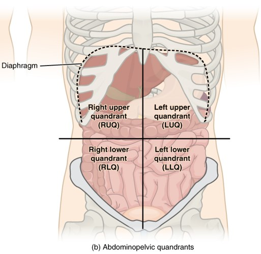
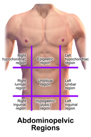
1. Anatomical Regions; Quadrants
2. Compare/contrast anatomy vs physiology
Anatomy - study of structures in the body (cells, organs)
Physiology - study of function of bodily structures
3. General functions of all body systems
Nervous System:
Functions: Controls and coordinates body activities, processes sensory information, and enables communication between different parts of the body.
Muscular System:
Functions: Allows movement, provides support and posture, generates heat.
Skeletal System:
Functions: Provides structural support, protects internal organs, facilitates movement, stores minerals (such as calcium), and produces blood cells.
Circulatory System (Cardiovascular System):
Functions: Transports oxygen, nutrients, hormones, and waste products throughout the body; helps regulate body temperature.
Respiratory System:
Functions: Facilitates the exchange of gases (oxygen and carbon dioxide) between the body and the environment; helps regulate pH.
Digestive System:
Functions: Breaks down and absorbs nutrients from food; eliminates waste.
Excretory System (Urinary System):
Functions: Filters and eliminates waste products from the blood; regulates electrolyte and fluid balance.
Endocrine System:
Functions: Produces and secretes hormones that regulate various body functions, including metabolism, growth, and reproduction.
Reproductive System:
Functions: Facilitates reproduction and the continuation of the species.
Integumentary System (Skin):
Functions: Protects against physical injury, pathogens, and dehydration; helps regulate body temperature; houses sensory receptors.
Immune System:
Functions: Defends the body against pathogens (such as bacteria, viruses, and fungi); recognizes and eliminates abnormal cells (e.g., cancer cells).
Lymphatic System:
Functions: Maintains fluid balance, transports fats from the digestive system, and supports the immune system.
4. Elements of a control system
For maintaining homeostasis (steady internal conditions)
The receptor detects change
Via the AFFERENT pathway (a for approaching) information is sent to control center
Control center analyzes, and decides the proper response
The efferent pathway then has the info
The info travels along the efferent pathway to effector
Effector responds in the proper way, which results in the homeostasis level again
5. Negative and Positive feedback mechanisms; know examples
Negative feedback effects of a reaction slow or stop the reaction
An example of negative feedback is the regulation of the blood calcium level, parathyroid glands secrete a parathyroid hormone
Brings body back to homeostasis
positive feedback amplifies the difference between the reference input/output, makes it stable, can become unstable
An example of positive feedback is increasing to push something farther, blood clots, and contractions in pregnancy
6. Anatomical position; orientation and directional terms; cavities; sections/planes (cuts)
Anatomical position- front facing palm facing front
Anterior - front
Posterior - back
Superior - above
Inferior - below
Medial - midline of body
Lateral - away from midline (sides)
Proximal - (for limbs) close to point of attachment
Distal - (for limbs) - far from point of attachment
Superficial - shallow
Deep - internal
Sagittal section - split in left and right
median/midsagittal - equal left and right
frontal/coronal - anterior posterior (slice like bread)
Transverse - superior and inferior parts (cut in half like a banana)
Dorsal body cavity - back of body, head, and back of trunk
Cranial cavity - brain and protected by skull
Spinal cavity - has spinal cord and protected by the vertebrae
Ventral body cavity - thoracic cavity and abdomino pelvic cavity
Subdivision by diaphragm of two cavities (thoracic cavity and abdomino)
Up (superior region) - stomach, liver, pancreas, spleen, gallbladder, kidneys, small intestine, and most of large intestine
Down (inferior region) - reproductive organs, bladder, rectum
Thoracic cavity - heart, lungs, other organs
7. Acids and bases; pH
Acids have a pH < 7
Bases have a pH > 7
8. What is an inorganic compound; know examples
Inorganic compounds - substance where two or more chemical elements (other than carbon) are combined, in definite proportions
Small and simple molecules
Water, salt, calcium carbonate, baking soda, copper(II)oxide, chlorate, tin(IV)chloride, lead(II)nitrate, iron(II), hydrogen sulfide, phosphate, phosphoric acid, hydrofluoric acid, hydrogen cyanide, magnesium oxide,
9. All types of reactions (synthesis, decomposition, hydrolysis, neutralization, dehydration
Decomposition: breaking down of something (all the chem ppl better know this our chem finals are right after)
Exchange: synthesis AND decomposition occur, so its essentially A+BC > AB+C (thats lit single replacement)
Hydrolysis - basically water is taken as a reactant, so then the water breaks the large molecule into tiny molecules (or if you want technicalities: reactions with water where monomers are released by the addition of water molecules adding OH and H to another)
Dehydration: chemical reaction in which water molecule is eliminated from the reactant molecule
Neutralization: acid and base react to form water and salt
10. Functions of proteins
Structural support
50% of body’s dry weight
Build and repair tissues, building blocks of amino acids
Help in enzymes and chemical reactions, many proteins speed up chemical reactions
Transporting molecules, proteins help transport oxygen, blood, and lipids through the body
Immune system protections as antibodies are proteins that help immune system fight viruses and such
Hormones, proteins like insulin act as hormones that regulate various body functions, like blood sugar level
Hydrogen bonds for intramolecular bonding
11. ATP: structure, function, where it’s made & how it’s made when you need a little vs. a lot
Consists of three main components, sugar molecule called ribose, nitrogenous base called adenine, and chain of three phosphate groups
Phosphate groups are connected by energy bonds which store energy that can be broken and generate energy
ATP is made through cellular respirations, occurring in the mitochondria
Glucose is broken down in a series of chemical reactions which releases energy (LOTS of ATP)
Energy is used to convert ADP to ATP by adding phosphate group, which is known as phosphorylation (quick ATP)
ATP is made and used to provide energy for anything in our cells
Needed to carry out tasks like muscle contraction, protein synthesis, or active transport
12. Cell junctions
Specialized structures that connect cells together in tissues
Play a crucial role in maintaining the integrity and function of tissues
Tight junctions - junctions which seal the space between cells, form barriers that prevent substances from leaking between cells
Epithelial tissues, lining of digestive tract
Adherens junctions - junctions which help cells adhere to each other and provide mechanical strength to tissues
Formed by proteins called cadherins,maintain structure of tissues
Desmosomes: desmosomes are similar to spot welds that hold cells together
Provide strong connections, are found in tissues that experience mechanical stress (skin and heart)
Gap Junctions - allow direct communication between cells, allow small molecules and ions to pass through between cells, exchange of signals and coordination of cellular activities
13. Classification and characteristics of epithelial tissues
In epithelial tissues, the cells are squished together and most likely form sheets
Simple squamous epithelium - tissue with a single layer of flat scale like cells, found in areas where diffusion and filtration occur, (blood vessels, air sacs in lungs)
Stratified squamous epithelium - tissue with multiple layers of flat cells. Protection against mechanical stress, found in skin, mouth, and esophagus
Simple cuboidal epithelium - this tissue consists of a single layer of cube shaped cells. It’s involved in secretion and absorption and its in kidney tubules and glands
Simple columnar epithelium - this tissue has a single layer of tall, column-shaped cells. It’s involved in absorption and secretion and is found in the lining of the digestive tract
Pseudostratified columnar epithelium - this tissue is stratified but is actually a single layer of cells of varying heights. It’s found in the respiratory tract and has cilia that help move mucus and particles
Basically stratified = multiple layers, cuboidal = cube shape cells, squamous = flat cells, simple = one layer, columnar = tall cells
Protection from injury, pathogens, substances
Absorption - simple columnar epithelium, tissue which lines small intestine, specialized for absorbing nutrients from digested food and transporting in blood stream
Secretion - epithelial tissue is involved in the secretion of various substances, glands, produce + release substances like hormones, enzymes, and sweat
Sensation - some epithelial tissues are responsible for detecting and transmitting sensory information (taste buds)
Filtration and excretion - epithelial tissue in kidneys play role in filtering waste products from blood - excreted as urine
Apical surface - exposed to external environment or body cavity - absorbs secrets, protects,
Lining of small intestine - apical layer of epithelial cells - microvilli - tiny finger-like projections that increase the surface area for absorption
Basement membrane - attached to a thin, non-cellular layer called the basement membrane. Provides support and helps anchor the epithelial tissue to underlying connective tissue
Lower surface of epithelium, basal lamina, rest on basement membrane, consists of reticular connective tissue securing the upper layers
avascular + relies on circulatory system to obtain food + oxygen
Regenerates easy if well-nourished
One layer - absorption, secretion
Multiple - protection
14. Endocrine and exocrine glands
Exocrine glands - secrete products (enzymes, mucus, sweat, oil) into ducts which carry substances to a specific location on the body’s surface, or a body cavity
These can be sweat glands, salivary glands, and oil (sebaceous glands), they help with processes like cooling the body, lubricating surfaces, and aiding in digestion
Endocrine glands - release secretions, or hormones, directly into the bloodstream. Hormones travel throughout the body to target specific cells/organs, regulating bodily functions.
Examples include the pituitary gland, thyroid gland, adrenal gland
Maintain homeostasis, growth, metabolism, reproduction
15. Compare/contrast smooth, cardiac, skeletal muscles tissue
- Location: It's attached to bones.
- Function: It's responsible for voluntary body movements, like when you decide to move your arm or leg.
- Appearance: Skeletal muscle looks striated, meaning it has prominent cross-stripes.
- Other Names: Some folks call it voluntary muscle because we have control over it.
- Stimulation: It's stimulated by consciously directed nerve activity. Basically, when you want to move, your brain sends signals to activate the muscle.
- Structure: The individual cells of skeletal muscle are called muscle fibers. They have many nuclei and come in different lengths and diameters.
- Organization: The muscle fibers are highly organized, kinda like in a cool pattern.
- Cell Membrane: The cell membrane of a muscle fiber is called the sarcolemma.
- Connective Tissue: There are a few types of connective tissue associated with skeletal muscle:
- Endomysium: It's a fine protective sheath that covers the sarcolemma.
- Perimysium: This fibrous membrane bundles muscle fibers into fascicles, which are like little groups of muscle fibers.
- Epimysium: It's a thick connective tissue that encloses all the fascicles in a muscle.
- Connections: Skeletal muscle connects to bones, cartilage, or other connective tissue through a couple of structures:
- Tendon: It's like a cord that connects the muscle to bone, cartilage, or other connective tissue.
- Aponeurosis: This is a flat, sheetlike structure that also connects muscle to bone, cartilage, or other connective tissue.
- Overall Enclosure: Tendons and aponeuroses are what connect the skeletal muscle to the bones, cartilage, or other connective tissue.
- Characteristics: Smooth muscle is small and spindle-shaped. It doesn't have the cross-stripes you see in skeletal muscle, so it's nonstriated. Plus, it's involuntary, meaning we can't consciously control it. Smooth muscle cells have a single nucleus.
- Other Names: Some call it visceral muscle.
- Location: You can find smooth muscle in the walls of internal organs like the stomach, intestines, urinary bladder, and respiratory passages.
- Arrangement: The cells of smooth muscle are arranged in layers, both longitudinal and circular.
- Function: Smooth muscle helps with coordinated contractions. It alternately contracts and relaxes, which changes the size and shape of the organ it's in. For example, it helps move food through the digestive system, empties the bladder, and even changes the diameter of blood vessels.
- Control: The autonomic nervous system, which is part of our automatic bodily functions, controls smooth muscle.
- Peristalsis: Smooth muscle is also responsible for the wave-like squeezing of the digestive tract walls during digestion. It's called peristalsis.
:
- Location: Muscle in the heart is solely found in the walls of the heart.
- Characteristics: This muscle is branched, cross-striated (meaning it has stripes), and involuntary (we can't control it consciously).
- Control: It's under the autonomic nervous system, which means it's controlled automatically by our body.
- Arrangement: The cells in heart muscle form an interconnected network of figure-eight or spiral-shaped bundles at intercalated discs.
- Function: The main job of this muscle is to coordinate the contraction of neighboring cells, which produces the heartbeat.
16. Characteristics of nervous tissue and its supporting cells
Nerve cells = neuron - irritability, conductivity, electrochemical impulse
Cns - central nervous system - brain and spinal cord
Pns - peripheral nervous system - everything outside of the brain and spinal cord
Excitable - capable of sending and receiving electrochemical signals that provide the body with information
Astrocytes are common in the CNS - they brace and anchor neurons
Microglia are in the CNS and they protect by destroying threats
Ependymal are in the central nervous system - protect spinal cord
Oligodendrocyte - CNS - makes myelin sheath
Shwann cells - the PNS - makes myelin sheaths
Satellites - PNS - protect cell body of neuron
17. Where transitional epithelial tissue can be found
Lining the structures of the urinary system, ureters, urinary bladder, urethra
18. Classification and characteristics of connective tissues
Osseous tissue - support protect, movement, blood cell production, mineral storage, metabolism regulations, fat storage, acid-base, balance, hematopoiesis
BONE
Hyaline cartilage - joint support, shock absorption, respiratory support, growth plate formation, bone development, fetal skeleton, resilience/flexibility
Articular surfaces, coastal/nasal cartilage, respiratory tract, fetal skeleton growth epiphyseal plates
Fibrocartilage - joint stability, load bearing, spinal support, tendon/ligament attachments, resilience, shock absorption, adaptation to stress, injury repair
Elastic cartilage - maintaining shape, flexibility, resilience, vibrational damping, supporting structure, elasticity, protection
Blood- transportation of substances, immunity, clotting, regulation of body temperature, pH, electrolyte balance, blood pressure regulation, hydration, transport of gasses
Dense regular tissue - strength and tensile resistance
Dense irregular tissue - strength and resistance to tension in multiple directions
Loose - Areolar - support and structure, binding, packaging, immunity, nutrient, waste transport, phagocytosis, wound healing, nerve support
Loose - adipose - energy storage, insulation, protection, hormone regulation, body shape, wound healing, endocrine functions, reproduction, fertility
Loose - reticular - structure and support, lymphatic system support, blood cell formation, endocrine gland supply, filter, remove cell debris, immune function, transport/exchange, tissue repair
19. Cartilage; chondrocytes; regeneration; collagen fibers:scar tissue
Cartilage is a firm and flexible connective tissue that is found in various parts of our body, such as our ears, nose, and joints.
Chondrocytes are the specialized cells found in cartilage. They are responsible for producing and maintaining the extracellular matrix of the cartilage, which is made up of collagen fibers and other components.
One interesting characteristic of cartilage is its limited ability to regenerate. Unlike other tissues in our body, cartilage has a low blood supply, which makes it harder for it to heal and regenerate on its own. However, there are some techniques being researched and developed to help promote cartilage regeneration, such as tissue engineering and stem cell therapy.
When cartilage is damaged, the body's natural response is to form scar tissue. Scar tissue is composed of collagen fibers that help to repair the injured area. However, scar tissue is not as strong or flexible as healthy cartilage, which can sometimes lead to long-term issues.
20. Vascular vs. avascular
Vascular = blood
Avascular = no blood
21. Cell structures that function to provide mobility
Flagella - long, whip like structures which extend from the cell and move in a wave-like motion
Found on cells like sperm cells, helping them to swin and navigate through fluids
Cilia are shorter, more numerous - tiny hairs that cover the surface of certain cells
Work together in coordinated movements - create a wave like motion, helps move substances along the surface of the cell
-found in various places, respiratory tract, help move mucus and trapped particles
Microvilli - tiny, finger-like projections that extend from the surface of certain cells, in tissues that are involved in absorption
Increase the surface area of the cell, allows for more efficient absorption of nutrients and other substances
Involved in absorption of nutrients/other substances
Involved in absorption of nutrients from the food we eat, the reabsorption of water/electrolytes in kidneys
Little extensions that help cells maximize their absorption capabilities
22. Peristalsis
Involuntary constriction and relaxation of the muscles in the intestine or another canal, creating wave like movements that push the contents of the canal forward
23. Epithelial and connective tissue membranes: cutaneous, mucous, serous, synovial
Cutaneous - skin, largest organ in our body, composed of stratified squamous epithelial tissue, underlying connective tissue
Protects our body from external elements and helps regulate temperature
Mucous membrane - lines various body cavities and organs that are open to the exterior, respiratory, digestive, reproductive tracts, consist of epithelial tissue and underlying connective tissue
Secrete mucous
Serous membrane - line internal cavities of the body and cover organs within the cavities,consist of a layer of simple squamous epithelium, thin layer of connective tissue
Secrete a watery fluid that helps reduce friction between organs during movement
Synovial membrane - line the joint cavities and produce synovial fluid, which lubricates the joints and reduces friction during movement, composed of connective tissue/do not have epithelial layer like other three membranes
24. Functions of the integumentary system
Body temperature regulation, cell fluid maintenance, synthesis of Vitamin D, detection of stiimuli
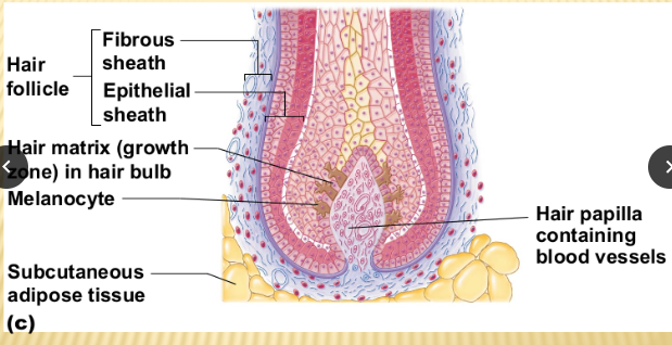
25. Structure of the hair
Outer tissue - connective, inner is epithelial
Root is enclosed in follicle, an d the shaft, or the dead part coming out, comes out
The shaft is hard kertinized epithelial cells
The melanocytes provide pigment for hair color
Hair is from hair bulb in STRATUM BASALE
26. Proteins of the integumentary system: keratin, melanin, carotene, hemoglobin
Keratin - major component of hair, nails, and outer layer of skin
Provides strength and protection to these structures, helping to keep them durable/resistant to damage
Melanin - pigment that gives color to our skin/hair/eyes - produced by specilized cells called melanocytes - amount and type determines skin tone/protects skin from harmful effects
Eumelanin - dark
Pheomelanin - light
Carotene - pigment that gives fruits orange color, when consuming foods rich in carotene, body converts it to vitamin A, important for maintaining healthy skin/vision
Hemoglobin - not protein of integ system - hemoglobin is protein found in red blood cells that carry oxygen throughout our body
Gives our blood red color - can be seen through the skin, like lips/cheeks
27. Glands of the skin: locations and functions
Sebaceous glands - found all over skin - except for palms of hands/soles of feet, secrete oily substance called sebum, keeps our skin from drying out
Particularly active during adolescence - oily skin/acne
Sweat glands
Eccrine glands - glands are found all over our body and are responsible for regulating body temperature
Produce sweat, cools down body when it evaporates from skins surface
Apocrine glands - glands found in armpits/genital area, active during puberty, produce thicker sweat that can be broken down by bacteria = body odor
Ceruminous glands - glands are located in ear canal - produce cerumen, or earwax, helps protect the ear canal by trapping dust, debris, and bacteria
Mammary glands - only to females, produce milk during lactation, located in breasts, crucial role in nourishing infants
28. Epidermis: structure (layers) and their order
Stratum basale - bottommost layer of epidermis, contains cells called basal cells that continuously divide and produce new skin cells
Stratum spinosum - above basal layer, spiny appearance of cells, layer provides strength and support to skin
Stratum granulosum - consists of granular cells that contain keratin - protein which provides strength/waterproofing
Stratum lucidum - not always present in areas of the body, found in thick skin, llike palms of hands and soles of feet, translucent cells that protect skin
Stratum corneum - outermost layer, composed of dead skin cells that are constnatly shed and replaced, layer acts as a barrier, protecting the underlying layers, and preventing water loss
29. Dermis: all characteristics and their functions
Thickness and strength - dermis is thicker than epidermis and provides structural support, collagen and elastin fibers give skin strength, elasticity, flexibility
Blood vessels, dermis has lots, help nourish skin cells regulate body temperature
Play role in delivering oxygen/nutrients to skin
Nerve endings - dermis has lots of nerve endings - allow us to sense, touch, pressure, temperature, and pain
Hair follicles/glands - dermis houses hair follicles - responsible for for hair growth, contains sweat glands that help regulate body temperature by producing sweat, sebaceous glands produce sebum - keeps skin moist
Immune cells - dermis has immune cells - protect the skin from infections/foreign substances
30. Composition of nails
Nail plate - hard visible part of nail, covers nail bed, composed of layers of kertinized cells
Nail bed - skin under the nail plate - support/nourishment to nail plate, contains blood vessels and nerves - pink color
Lunula - whitish, crrescent-shaped area at base of nnail, visible part of nail matrix - responsible for nail growth
Cuticle - thin layer of skin that overlaps base of nail plate, protective barrier, preventing backteria/debris from nail bed
31. ABCD rule
A - asymmetry - mole/spot is asymmetrical
B - border - irregular, blurry, jagged = warning
C - color - color of mole, different shades of brown, black, red - problem
D - diameter - melanomas are typicalles larger than 6 millimeters
32. Characteristics of melanoma
Melanoma is a type of skin cancer that can develop from abnormal growth of pigment-producing cells called melanocytes. Here are some characteristics to be aware of when it comes to melanoma:
1. Asymmetry: Melanomas often have an irregular shape, where one half is different from the other half.
2. Border: The borders of melanomas may be uneven, jagged, or poorly defined, rather than smooth and round.
3. Color: Melanomas can have a variety of colors within the same lesion, including shades of brown, black, blue, red, or white.
4. Diameter: While melanomas are typically larger than 6 millimeters (about the size of a pencil eraser), they can also be smaller in size.
5. Evolution: Pay attention to any changes in the size, shape, color, or elevation of a mole or spot. Melanomas may evolve and exhibit these changes over time.
33. Structure/function of the Osteon (Haversian system); know this in great detail
Sure, Hritika! Let's dive into the structure and function of the osteon, also known as the Haversian system, in great detail.
The osteon is the basic structural unit of compact bone and plays a vital role in the support and strength of our skeletal system. It consists of several components that work together harmoniously.
At the center of the osteon, we have the central canal, also called the Haversian canal. This canal houses blood vessels, nerves, and connective tissue, providing nourishment and oxygen to the bone cells.
Surrounding the central canal are concentric layers of bone matrix called lamellae. These lamellae are composed of collagen fibers and mineralized calcium phosphate, giving bone its strength and resilience.
Between the lamellae, we find small spaces called lacunae. These lacunae house osteocytes, which are mature bone cells responsible for maintaining the bone tissue. Osteocytes communicate with each other and with the central canal through tiny channels called canaliculi, allowing for the exchange of nutrients and waste products.
Connecting neighboring osteons are perforating canals, also known as Volkmann's canals. These canals contain blood vessels and nerves that connect with the central canals, ensuring a continuous supply of nutrients and oxygen throughout the bone.
The osteon's structure allows for efficient nutrient and waste exchange, as well as the transmission of forces and mechanical stress. It provides strength, stability, and resilience to our bones, enabling them to withstand everyday activities and support our body.
34. Ossification
Process of bone formation
35. Foramen Magnum
A large opening at the base of the skull
Located at the back, where the spinal accord connects to the brain
Spinal cord passes through foramen magnum, allow3ng comms between brain and body
36. All of the bones (from your test diagram); know axial vs. appendicular skeleton too
The human body is made up of 206 bones, which provide support, protection, and help with movement. These bones can be categorized into two main groups: the axial skeleton and the appendicular skeleton.
The axial skeleton includes the bones along the central axis of the body. It consists of the skull, vertebral column (including the spine), and the ribcage. The skull protects the brain, while the vertebral column supports the body and protects the spinal cord. The ribcage encloses and protects the vital organs in the chest, such as the heart and lungs.
On the other hand, the appendicular skeleton includes the bones of the limbs and their attachment points. It consists of the arms, legs, shoulder girdle, and pelvic girdle. The arms consist of the humerus (upper arm bone), radius, and ulna (forearm bones), as well as the bones of the hand. The legs consist of the femur (thigh bone), tibia, and fibula (lower leg bones), as well as the bones of the foot. The shoulder girdle includes the clavicle (collarbone) and scapula (shoulder blade), while the pelvic girdle consists of the hip bones.
37. Articulations; joints
Articulations - location where two or more bones meet
38. Minerals stored in bones
Calcium, phosphorus, collagen
39. Hematopoesis
Process of creating a wide variety of blood and bone marrow cells
40. Anatomy of the long bone
Two parts
Diaphysis - epiphysis
Diaphysis - tubular shaft that runs between the proximal and distal ends of bone
Hollow region in diaphysis - medullary cavity - yellow marrow
Walls of diaphysis are composed of dense and hard - compact bone
Wide section end of bone - epiphysis - spongy bone
Red marrow in spaces of spongy bone
Epiphysis meets diaphysis at the metaphysis - narrow area that contains the epiphyseal plate - growth plate, layer of hyaline cartilage in growing bone
Medullary cavity - delicate membranous lining called the endosteum - bone growth, repair, remodeling
Outer surface of bone is covered with a fibrous membrane - periosteum
Periosteum contains - blood vessels, nerves, lymphatic vessels that nourish compact bone
Tendons + ligaments also attach to bones at the periosteum
Covers everything but the surface where epiphyses meet other bones
In this region - articular cartilage, thin layer of cartilage to reduce friction, act as shock absorption
41. Epiphyseal plate vs. line; location; function
-epiphyseal plate and line are found in long bones, like the ones in our arms and legs
-related to bone growth/development
-epiphyseal plate growth - located at ends of long bones
-made of cartilage
-responsible for bone lengthening during growth
-cells in epiphyseal plate divide and replace cartilage with bone - increase in bone length
-on the other hand, the epiphyseal line is formed when epiphyseal plate stops growing/ossifies - becomes bone
-marks location where growth plate used to be
42. Parathyroid hormone and what it does
The parathyroid hormone is produced by parathyroid glands - small glands near the thyroid gland
Regulates the levels of calcium and phosphorus in the body
Blood calcium is low = PTH in the bloodstream, PTH acts oon bones, kidneys, intestines to increase calcium, stimulating release of calcium from bones, enhancing the reabsorption of calcium by kidneys, promotes absorption of calcium from intestines
By increasing blood calcium levels, pTH maintins calcium balance, essential for various bodily functions, bone health, nerve function, muscle contractions
43. Function of specific bone markings
Processes and projections on bones - give attachment points for muscles/tendons/ligaments
Movement and stability
Depressions and openings in bones can serve as passageways for blood vessels and nerves, ensuring proper circulation and innervation
Articulating surfaces on bones form joints enabling smooth movement/flexibility
Fossae and cavities in bones - house and protect vital organs - like brain within the cranial cavity or heart within thoracic cavity
44. All bone cells: “osteo____”
osteoblasts - bone formation, produce/secrete the proteins and minerals that make up the bone matrix, crucial for bone growth/repair
Osteocytes - osteocytes are mature bone cells that are embedded in bone matrix, maintain and monitor bone tissue, play a role in regulating bone density and mineral homeostasis. Help in detecting mechanical stress on the bone and can trigger remodeling processes to strengthen the bone.
Osteoclasts - cells are involved in bone resorption, process of breaking down and removing old/damaged bone tissue, osteoclasts secrete enzymes, dissolve mineralized matrix, removal of bone tissue
Crucial for bone remodeling/ balance between bone formation and resorption
45. Stages of bone fracture healing
1. Fracture hematoma: Immediately after the fracture occurs, blood vessels at the site of the break rupture, leading to the formation of a blood clot called a hematoma. This hematoma provides a foundation for the next stages of healing.
2. Inflammatory phase: In this phase, the body's immune response kicks in. White blood cells and other inflammatory cells arrive at the fracture site to remove any debris and prevent infection. The damaged blood vessels start to regrow, and new blood vessels form to supply oxygen and nutrients to the healing bone.
3. Soft callus formation: During this stage, specialized cells called chondroblasts produce a soft callus made of cartilage. This callus acts as a bridge between the broken bone ends and helps stabilize the fracture. It's like a temporary scaffolding.
4. Hard callus formation: Over time, the soft callus is gradually replaced by a hard callus made of spongy bone. Osteoblasts, which are bone-forming cells, lay down new bone tissue to strengthen the fracture site. This process can take several weeks to months.
5. Remodeling: In the final stage, the hard callus is remodeled and reshaped to resemble the original bone structure. Osteoclasts, which are bone-resorbing cells, remove excess bone tissue, while osteoblasts continue to build new bone. This remodeling process can take months to years, depending on the severity of the fracture.
46. Types of fractures
Comminuted - bone breaks into many fragments - common in older people, bones are more brittle
Compression - bone is crushed - common in porous bones (osteoporotic bones)
Depressed - broken bone portion a is pressed inward - typical of skull fracture
Impacted - broken bone ends are forced into each other - commonly occurs when someone attempts to break a fall with outstretched arms
Spiral - ragged break occurs when excessive twisting forces are applied to a bone - common sports fracture
Greenstick - bone breaks incompletely, much in the way a green twig breaks - common in children, whose bones are more flexible than those of adults
47. Fontanels- location/function
-soft spots on baby’s skull, bones have not fully fused together, located on top, back, sides of the head
-functions of fontanels - to allow for the growth and flexibility of the baby’s skull during birth and early development
-close as the baby’s skull bones grow/fuse together
48. Uniqueness of hyoid bone
Only bone in the body which does not connect to any other bone
Is suspended in the neck by muscles and ligaments
-crucial role in supporting the tongue and aids in swallowing + speech
49. Unique features of cervical vertebrae; atlas and axis and their unique features
-cervical vertebrae, including the atlas/axis
-atlas (c1v) first one in the neck, supports weight of the head
Does not have a body like other vertebrae
Ring like structure for head movement
- axis (c2v) - second one in neck with special feature
dens / odontoid process
Bony projection that sticks up from body of axis, acts as pivot point for rotation of atlas and skull
Knob that allows head to turn laterally
50. Sternum; structure
Manubrium - broadest part of the sternum, shaped like a trapezoid/articulates with clavicles + first rib
Body - gladiolus - middle/longest part of the sternum. Relatively flat, connects to the manubrium process above, xiphoid below
Xiphoid process - smallest and lowest part of the sternum, small, cartilaginous structure which hardens with age
51. Arrangement of the vertebral column
Thoracic region - middle part and consists of the twelve thoracic vertebrae - connect to ribs and form chest
Lumbar region - lumbar region is the lower part of the vertebral column - consists of five lumbar vertebrae, which are larger to support the body
Sacral region - belo the lumbar with five fused vertebrae, form the sacrum, which connects spine to pelvis
Coccygeal region - lowest part, has coccyx which is known as the tailbone, made of three to five fused vertebraeccq1
52. Types and respective movements of each of the synovial joints
Ball-and-Socket join - shoulder, hip
Saddle joint - metacarpals to carpal at pollex
Condylar joint - metacarppals to phalanges
One bone end is round - convex,
One bone end is depressed (concave)
Pivot joint - C1/C2, proximal radius and ulna
Hinge joint - elbow, ankle, knee
Plane joint - gliding movements (wrist)
53. Skeletal muscle activity: special functional properties
1. Excitability: Skeletal muscles have the ability to respond to stimuli, such as nerve signals, by generating electrical impulses. This allows them to initiate muscle contractions.
2. Contractility: One of the key functions of skeletal muscles is their ability to contract or shorten. When stimulated, muscle fibers slide past each other, causing the muscle to contract and generate force.
3. Extensibility: Skeletal muscles can be stretched or extended without damage. This property allows for flexibility and movement in our joints, enabling us to perform a wide range of motions.
4. Elasticity: After being stretched, skeletal muscles have the ability to return to their original shape and length. This elasticity helps muscles maintain their structure and function, allowing for efficient movement.
5. Motor Unit Recruitment: Skeletal muscles are composed of motor units, which consist of a motor neuron and the muscle fibers it innervates. The recruitment of motor units allows for graded muscle contractions, enabling us to control the force and precision of our movements.
These special functional properties of skeletal muscle activity work together to facilitate our everyday movements, from walking and running to lifting and even facial expressions. Our muscles are truly remarkable!
54. What makes up a motor unit
A motor unit is composed of two main components: a motor neuron and the muscle fibers it innervates. The motor neuron is a specialized nerve cell that transmits signals from the central nervous system to the muscle fibers. These signals, known as action potentials, stimulate the muscle fibers to contract.
The motor neuron has a cell body located in the spinal cord or brainstem, and it extends its long, thread-like projection called an axon to reach the muscle fibers. The axon branches out into smaller branches called axon terminals, which make contact with individual muscle fibers.
Each motor neuron can innervate multiple muscle fibers, but each muscle fiber is innervated by only one motor neuron. The number of muscle fibers innervated by a single motor neuron can vary depending on the precision and control required for a specific muscle. For example, fine motor control muscles, like those in the fingers, may have fewer muscle fibers per motor neuron compared to larger muscles like the quadriceps.
When the motor neuron receives a signal from the central nervous system, it releases a neurotransmitter called acetylcholine into the neuromuscular junction, which is the point of contact between the motor neuron and the muscle fiber. Acetylcholine binds to receptors on the muscle fiber, triggering a cascade of events that lead to muscle contraction.
The coordinated activation of motor units, with varying numbers of muscle fibers contracting, allows for precise control and modulation of muscle force during movements.
55. How cross bridges are formed
Ah, cross bridges! They play a crucial role in muscle contraction. When a muscle receives a signal to contract, the myosin heads, which are part of the thick filaments in the muscle fibers, form cross bridges with the actin filaments, which are part of the thin filaments.
These cross bridges are formed when the myosin heads bind to specific binding sites on the actin filaments. This binding occurs due to the release of stored energy in the myosin heads, which causes them to change their shape and reach out to attach to the actin filaments.
Once the cross bridges are formed, a series of events take place. The myosin heads undergo a power stroke, pulling the actin filaments closer together. This results in the shortening of the muscle fiber and the generation of force.
After the power stroke, the myosin heads release from the actin filaments and reset to their original position. This allows for another round of cross bridge formation and subsequent muscle contractions.
The formation and cycling of cross bridges are essential for the sliding filament theory of muscle contraction, which explains how muscle fibers contract and generate force.
56. Details of the neuromuscular junction and the synaptic cleft
The neuromuscular junction is the point where a motor neuron and a muscle fiber meet. It's like a meeting point for the nervous system and the muscular system. When a nerve impulse reaches the end of a motor neuron, it triggers the release of a chemical called acetylcholine into the synaptic cleft.
Now, the synaptic cleft is the tiny gap between the motor neuron and the muscle fiber. It's like a little space that they have to bridge. When acetylcholine is released into the synaptic cleft, it diffuses across this gap and binds to receptors on the muscle fiber.
This binding of acetylcholine to the receptors triggers a series of events that ultimately lead to the contraction of the muscle fiber. It's like a signal that tells the muscle to do its thing!
After the acetylcholine has done its job, it gets broken down by an enzyme called acetylcholinesterase to prevent continuous muscle contraction. This allows the muscle fiber to relax and be ready for the next nerve impulse.
So, the neuromuscular junction and the synaptic cleft are important players in the communication between nerves and muscles. They ensure that the signals from the nervous system are transmitted to the muscles, allowing us to move and perform all sorts of amazing actions!
57. Interactions of skeletal muscles in the body (antagonists, etc…)
When it comes to the interactions of skeletal muscles in the body, there are a few key terms to know. One important concept is the idea of agonist and antagonist muscles.
Agonist muscles are the ones primarily responsible for producing a specific movement. They contract and generate force to create the desired action. For example, when you bend your arm, the biceps brachii muscle acts as the agonist.
On the other hand, antagonist muscles work in opposition to the agonist muscles. They relax and lengthen to allow the desired movement to occur smoothly. In the case of bending your arm, the triceps brachii muscle acts as the antagonist, as it relaxes to allow the biceps to contract and bend the arm.
This agonist-antagonist relationship allows for precise control and coordination of movements in the body. By working together, these muscles help create smooth and controlled actions.
There are also synergist muscles, which assist the agonist muscles in performing a movement. They help stabilize the joint or assist in the action. For example, when you lift a heavy object, your biceps brachii is the agonist, but muscles like the brachialis and brachioradialis act as synergists to assist in the movement.
Understanding these muscle interactions can give us insight into how our body moves and functions. It's pretty amazing how everything works together!
58. Muscle movements and which muscles can do which movements
1. Flexion: This movement involves bending or decreasing the angle between two body parts. Muscles like the biceps brachii in the upper arm and the hamstrings in the thigh are responsible for flexion.
2. Extension: The opposite of flexion, extension involves straightening or increasing the angle between two body parts. Muscles such as the triceps brachii in the upper arm and the quadriceps in the thigh perform extension.
3. Abduction: This movement refers to moving a body part away from the midline of the body. Muscles like the deltoids in the shoulder and the gluteus medius in the hip are responsible for abduction.
4. Adduction: The opposite of abduction, adduction involves moving a body part toward the midline of the body. Muscles such as the adductor muscles in the thigh and the pectoralis major in the chest perform adduction.
5. Rotation: This movement involves turning or twisting a body part around its axis. Muscles like the external obliques in the abdomen and the rotator cuff muscles in the shoulder perform rotation.
1. Flexion of the forearm: This movement involves bending the forearm towards the upper arm. The biceps brachii muscle is primarily responsible for flexing the forearm.
2. Plantar flexion: This movement refers to pointing the foot downward, like when you stand on your tiptoes. The gastrocnemius and soleus muscles in the calf perform plantar flexion.
3. Dorsiflexion: The opposite of plantar flexion, dorsiflexion involves pulling the foot upward, as if you're trying to touch your toes to your shin. The tibialis anterior muscle in the front of the lower leg is responsible for dorsiflexion.
4. Pronation: This movement involves turning the palm of the hand downward or facing backward. The pronator teres and pronator quadratus muscles in the forearm are responsible for pronation.
5. Supination: The opposite of pronation, supination involves turning the palm of the hand upward or facing forward. The biceps brachii and supinator muscles in the forearm perform supination.
59. Microscopic anatomy of skeletal muscle; know all of the parts down to the myofilaments
At a microscopic level, skeletal muscle is composed of several key components. The muscle cell itself is called a muscle fiber, which is a long, cylindrical structure. Inside the muscle fiber, there are smaller units called myofibrils.
Myofibrils are made up of repeating units called sarcomeres, which are the functional units of muscle contraction. Sarcomeres consist of thick and thin protein filaments called myofilaments.
The thick filaments are made up of a protein called myosin, while the thin filaments are primarily composed of actin, along with other regulatory proteins like troponin and tropomyosin. These myofilaments are arranged in a highly organized pattern within the sarcomere.
When a muscle contracts, the myosin heads on the thick filaments interact with the actin filaments, causing them to slide past each other. This sliding action is what leads to muscle shortening and contraction.
So, to recap, skeletal muscle is made up of muscle fibers, which contain myofibrils. Within the myofibrils, we find sarcomeres composed of thick myosin filaments and thin actin filaments, among other proteins.
60. The sliding filament theory- what it does, how it works; what is shortening
Imagine your muscles are made up of tiny units called sarcomeres. Inside each sarcomere, there are two types of protein filaments: thick filaments made of myosin and thin filaments made of actin.
When a muscle contracts, the myosin heads on the thick filaments grab onto the actin filaments and pull them towards the center of the sarcomere. This action is like a bunch of tiny muscle fibers sliding past each other, hence the name "sliding filament theory."
As the myosin heads pull the actin filaments, the sarcomeres shorten. This shortening of sarcomeres is what we refer to as muscle contraction. It's like when you flex your bicep and it becomes shorter and thicker.
The sliding filament theory is a simplified way to understand the complex molecular interactions that occur during muscle contraction. It's pretty amazing how our muscles work!
61. Neurotransmitters and how they are released; which one causes skeletal muscle contraction
Neurotransmitters are like messengers in our bodies, helping to transmit signals between nerve cells. When a nerve impulse reaches the end of a neuron, it triggers the release of neurotransmitters into the synapse, which is the tiny gap between the sending neuron and the receiving neuron.
The release of neurotransmitters is a fascinating process. Once the nerve impulse reaches the end of the neuron, it causes tiny sacs called synaptic vesicles to fuse with the neuron's cell membrane. These vesicles contain the neurotransmitters. When the vesicles fuse with the membrane, they release the neurotransmitters into the synapse.
Once released, the neurotransmitters travel across the synapse and bind to specific receptors on the receiving neuron. This binding process helps transmit the signal from one neuron to another.
Now, when it comes to skeletal muscle contraction, the neurotransmitter responsible for initiating muscle contractions is acetylcholine (ACh). ACh is released from nerve endings onto muscle fibers and binds to receptors on the muscle cell membrane. This binding triggers a series of events that ultimately leads to muscle contraction.
62. The role of creatine phosphate and how it makes ATP
Here's how it works: Creatine phosphate stores high-energy phosphate groups. When our muscles need ATP (adenosine triphosphate) for energy, creatine phosphate donates one of its phosphate groups to ADP (adenosine diphosphate), converting it back into ATP.
This process is called phosphorylation. By transferring its phosphate group to ADP, creatine phosphate helps regenerate ATP, which is the primary energy source for muscle contractions.
Think of it as a backup generator for ATP production. It allows our muscles to maintain their energy levels during short bursts of intense activity, like lifting heavy weights or sprinting.
63. Why skeletal muscle fatigue occurs
Depletion of energy sources, like ATP because of too much muscle activity
Accumulation of metabolic byproducts: as the muscles work, produce metabolic byproducts like lactic acid, if not cleared build up, muscle fatigue occurs
Impaired muscle function: prolonged muscle contraction impairs the ability of muscle fibers to contract efficiently, reduced force production and muscle fatigue
electrolyte imbalance: intense exercise can lead to loss of electrolytes (sodium, potassium, calcium) through swear, play crucial roles in muscle contractions and their imbalances lead to muscle fatigue
64. Isometric vs. isotonic contraction
Isometric contractions: in isometric contractions, the muscle contracts without change in length of the muscle
Isometric contractions maintain posture and stability
Isotonic contractions: the muscle contracts and changes in length, like curling a weight
Walking, running, lifting
65. Anaerobic glycolysis: when it occurs and how it makes ATP
66. What is atrophy
When a muscle or tissue shrinks/decreases in size.
Disuse, aging, medical condtions
When muscle is not used or stimulated enough, loses strength and mass