


3.8: Nervous system
Components
The nervous system detects changes in stimuli (inside or outside of the body), processes and stores information and initiates responses, which happens in 5 main steps:
Stimulus - This is a detectable change in the internal or external environment which will eventually produce a response.
Receptor - These are specialised sensory cells, such as pressure sensors in skin and complex sense organs such as the ear and the eye.
They are transducers, as energy is detected in one form and converted to electrical energy.
These impulses travel along neurons and are called nervous impulses.
Coordinator - Signals to effector to initiate a response.
Effector - Initiates a response, can either be a muscle or gland.
Response - Alteration in how the body acts in response to the stimulus.
Nervous system
There are two main parts:
The central nervous system (CNS) comprises the brain and spinal cord.
It processes information provided by a stimulus.
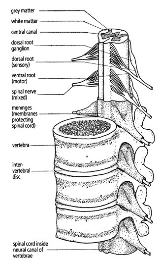
Both the brain and spinal cord are protected by tough, protective membranes known as meninges.
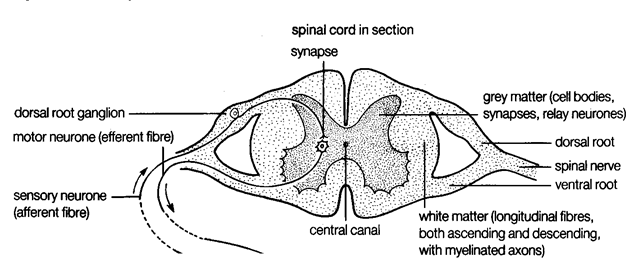
The spinal cord has many parts:
The central canal is the centre of the cord, filled with cerebral spinal fluid.
Surrounding it is grey matter, which is largely made up of the nerve fibres of relay neurons and the cell bodies of relay and motor neurons, causing it to be darker in colour (due to the nuclei in these cell bodies).
Surrounding this is white matter, which contains nerve fibres surrounded by fatty myelin causing it to be lighter in colour.
On either side of this are the two roots, which divide from the spinal nerve:
The dorsal root is on top, and contains a sensory neuron. It’s body causes a swell in the dorsal root known as the dorsal root ganglion. Electrical impulses move into the spinal cord through it.
The ventral root is on the bottom, and contains a motor neuron. Electrical impulses leave the spinal cord through it.
Around this is the protective layer - the meninge.
This is surrounded by a vertebra - a bone.
In between each vertebra is an inter-vertal disc, which prevents them from rubbing against each other.
The peripheral nervous system (PNS) uses the peripheral nerves (nerves outside of the spinal cord) and comprises 2 parts:
The somatic nervous system - Pairs of nerves that originate in the brain or the spinal cord, and their branches.
These nerves contain fibres of sensory neurons, and carry impulses from receptors to the CNS, and motor neurons, which carry impulses away from the CNS to effectors.
Helps the body process sensory information, reflex arcs and voluntary movements.
The autonomic nervous system - Provides unconscious control of the functions of internal organs, such as digestion.
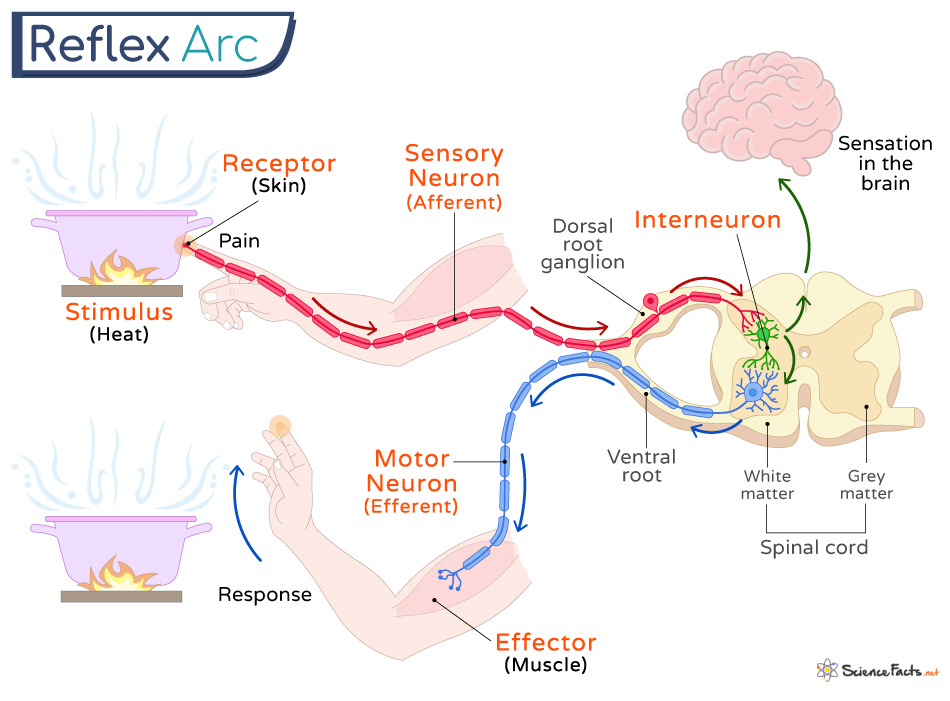
Reflex arc
This is the simplest type of response to a stimulus.
It is the neural pathway taken by the nervous impulses of a reflex action.
It is rapid, automatic and generally protective in purpose.
Decision making areas of the brain are not involved, and the action is involuntary.
An example using the withdrawal reflex:
A hot pan is touched.
This is detected by sensory receptors in the skin.
They pass the impulse to sensory neurons up the arm, which send it to the spinal cord.
A relay neuron in the spinal cord transmits the impulse from a sensory neuron to a motor neuron.
The motor neuron sends the impulse to an effector, which is a muscle.
This brings about the response; the arm muscles contract and the hand is removed from the heat source.
Nerve nets
Animals in the early fossil record do not have nervous systems.
Later in the fossil record are animals with radial symmetry with a nerve net nervous system.
Even later animals have bilateral symmetry and CNS, similar to humans.
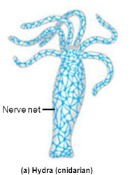
Nerve nets are the simplest type of nervous system, comprising a diffuse network of cells that group into ganglia without forming a brain. There are two cell types:
Ganglion cells which provide connection in many directions.
Sensory cells which detect stimuli.
An example is Hydra, which has a simple patterned nerve net which is easy to manipulate and regenerates rapidly.
The nerve net of Hydra is located in the ectoderm - the outer of it’s two layers of cells. This allows it to sense light, physical contact and chemicals.
It can contract, perform locomotion (move from one place to another), hunt and feed using this information. However, conduction speed is slowed.
Yet it cannot detect the direction of stimuli, only the size.
Neurons
Types
There are three main types, in order of occurrence:
Sensory neurons carry impulses from sense receptors/organs into the CNS.
Relay neurons receive impulses from sensory neurons or other relay neurons, and then transmit them to motor neurons or other relay neurons.
Motor neurons carry impulses from the CNS to effector organs; muscles or glands.
Structure
The cell body contains the nucleus and granular cytoplasm (contains lots of ribosomes). There are also Nissl granules, which are cytoplasmic granules comprising ribosomes grouped on RER.
In the sensory neuron, it is to the side of the neuron, attached via myelin sheath. In the relay neuron, this is in the middle of the neuron, between segments of myelin sheath. In the motor neuron, this is located in the dendrites.
Dendrites are thin fibres which carry impulses towards the cell body, and neurons can have several. They have a large SA to increase information received. This is at the top of the neuron.
Axon is a thin fibre that carries impulses away from the cell body. There is only one, and it runs throughout the cell body.
The myelin sheath speeds up electrical transmissions while protecting nerve fibres. It covers the entire axon aside from the dendrites and axon terminal. There are two parts:
Schwann cells. These surround and protect nerve fibres. They form in embryos, wrapping around 20-30 around the developing axons and then withdraw cytoplasm, leaving a multi-layer myelin sheath containing many phospholipids - which are good insulators. Their nuclei are on the outside, facing away from the axon.
Nodes of Ranvier. These are 1um gaps in myelin sheath where the adjacent Schwann cells meet, and the axon membrane is exposed. This allows impulses to be transmitted rapidly.
Axon terminals are located at the end of the neuron, and secrete neurotransmitters to adjacent neurons.
Have synaptic end bulbs, which are seen as swellings. This is where neurotransmitters are synthesised.

Nervous impulse
Resting potential
Neurons are excitable cells, meaning they can change their resting potential (the potential difference across the membrane of a cell when no nervous impulse is being conducted.)
This is not a common trait of cells.
The potential difference (differences in electrical charge between the inside of an axon and the outer tissue fluid) across neuron cell membranes is around 70 mV.
Both inside and outside the membrane are positively charged, however due to this difference the resting potential is -70 mV.
This potential difference across cell membranes means it is described as polarised.
This is due to two things; the negative ions of large proteins, organic acids (e.g pyruvate) and organic phosphates (e.g ATP4-) and the uneven distribution of inorganic ions.
There are two processes of uneven distribution, using transport protein:
Active ion transport - Axon’s actively transport two ions, K+ and Na+, against their concentrations gradients.
This is done using sodium-potassium exchange pumps, which are transmembrane proteins that bring K+ ions in and pump out Na+ ions.
They have ATPase activity.
For every 3 Na+ ions pumped out, only 2 K+ ions are brought in, contributing to the uneven distribution.
Facilitated diffusion - The active transport causes a high K+ concentration inside the axon, and a high NA+ concentration in the tissue fluid outside the axon. This leads to facilitated diffusion occurring.
However, the membrane is selectively permeable, and is about 100x more permeable to K+ ions.
This is due to the channels that diffuse the ions being able to open and close in response to voltages across the membrane (known as voltage-gated), and during resting potential most of the Na+ channels are closed, while only some K+ channels are.
Therefore, Na+ ions are pumped out faster than K+ ions enter, causing the -70 mV.

Action potential
Oscilloscopes can be used to show changes in voltage across membranes over time. They produce oscilloscope traces.
It works by placing microelectroduces in axons and the bathing solution surrounding it, measuring the voltage changes, often using squid axons as they are around 0.5 mm in diameter.
It measures the magnitude and transmission speeds as well as the pattern of impulses generated in different parts of the nervous system and different situations.
These conclusions won a Nobel prize.
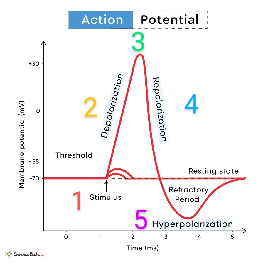
Below is an oscilloscope trace, which has 5 main stages:
1 - A stimulus generates energy, and once over the -55 mV threshold value the voltage-gated sodium channels in the axon membrane open.
2 - Depolarisation: An increasing amount of Na+ ions enter the axon via chemiosmosis, causing the positivity of the axon to increase. This opens more voltage-gated sodium channels, and therefore even more Na+ ions can enter.
Depolarisation is the temporary reversal of potential difference, where the inside is less negative than the outside as an action potential is transferred.
3 - The potential difference reaches +40 mV from -70 mV, which is the action potential.
4 - Repolarisation: Once this +40 mV is reached, the voltage-gated sodium channels close and K+ ions diffuse out via their concentration gradient. The positivity inside the membrane decreases.
5 - Hyperpolarisation - More K+ ions diffuse out than Na+ diffuse in, causing the potential difference to be more negative than resting potential.
The sodium-potassium exchange pump pumps K+ ions back in and Na+ ions back out, restoring the resting potential.
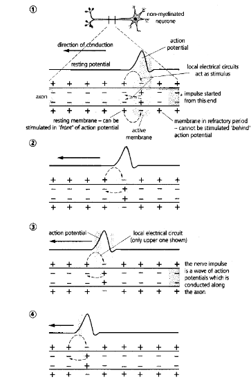
Movement along an axon
At the site of the action potential, the potential difference is higher than the rest of the neuron due to the movement of Na+.
This sets up local current, with Na+ moving laterally through the axon.
This depolarises adjacent sections of the membrane, opening more voltage-gated sodium channels in those regions.
The Na+ moves laterally again, repeating the process.
At the original action potential site, sodium channels are inactivated and unable to reopen until resting potential is reestablished.
This prevents a new action potential from being triggered, therefore preventing the action potential from moving backwards in the axon, keeping in in one direction.
This is the absolute refractory period.
For the next 5-10 ms, which is the hyperpolarisation phase, strong enough impulses can cause an action potential to start.
This occurs while sodium-potassium transfer pumps are still restoring resting potential.
This is known as the relative refractory period.
Nerve and impulse properties
‘All or nothing‘ law
If stimulus intensity does not reach -55 mV, no action potential is generated.
If initiated, however, it will always be the same size of +40 mV and always remains this size as no energy is lost in transmission.
Increases in stimulus intensity don’t increase action potential, but increase the frequency.
These rules - frequencies are either transmitted or not and are always the same size, is the all or nothing law.
This is done to allow action potential to act as a filter for minor stimuli, therefore not overloading the brain with nervous impulses.
Conduction speed
There are three major factors:
Temperature - At higher temperatures, there is more kinetic energy therefore ions are able to move faster.
Therefore, mammals and birds, the warm-blooded taxa, have faster moving impulses and higher reaction times than all other animal groups.
Axon diameter - The bigger the diameter, the less resistance, meaning more Na+ ions can enter, meaning depolarisation is faster and therefore faster impulse travel.
Non-myelinated human axons are around 0.2-1.5 um in diameter, which would only allow for slow impulse transmission.
Animals which live in lower temperatures, such as marine invertebrates including squid, compensate by having larger axons. Squids axons are up to 1 mm.
Earthworms, which live in warm environments, also have large axons - 700 um, which allows for rapid escape responses.
Myelination - Insulates the axon, therefore increasing transmission.
Myelinated nerve fibres only depolarise at areas of low resistance, which is the nodes of Ranvier, which is where the voltage-gated ion channels occur. This is therefore where Na+ ions enter.
This leads the action potential to seemingly jump from node to node, which is known as saltatory conduction.
It is rapid due to nodes of Ranvier being 1 mm apart.
Synapse
There are two types of synapse:
Electrical - the gap is 3nm across, small enough for an electrical impulse to be directly transmitted between neurons.
Chemical - the gap is 20 nm, which is too large for the impulse to jump.
It is the majority of synapses.
Axon branches lie close to dendrites of other neurons without touching, and the impulse is instead transmitted via neurotransmitters.
These chemicals diffuse across the synaptic cleft, from the pre-synaptic membrane of one neuron to the post-synaptic neuron of an adjacent neuron, which transmits the impulse.
They:
Transmit information between neurons.
Only pass information in one direction, maintaining the precision.
Act as junctions.
Protect the nervous system from overstimulation, as the impulse size is maintained.
Low-level stimuli is also filtered as it is only passed on if it reaches -55 mV.
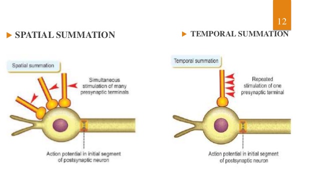
This can be built up to using:
Temporal summation - Where multiple action potentials are passed on, building up to reach the threshold value.
Spatial summation - Several pre-synaptic neurons contribute to the same post-synaptic neuron, building up to an action potential.
Structure
The pre-synaptic neuron is where the nervous impulse leaves.
The post-synaptic neuron is where the nervous impulse is entering.
Between them is the synaptic cleft.
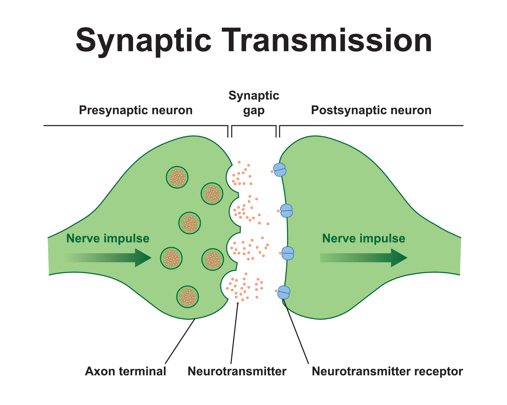
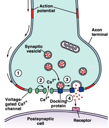
Transmission
There are 4 steps:
When an action potential reaches the pre-synaptic end bulb, the membrane permeability is altered, which opens voltage-dependent calcium channels.
These diffuse into the end of the bulb, down a concentration gradient from the synaptic cleft.
This influx of Ca+ causes synaptic vesicles in the pre-synaptic bulb to move towards and fuse with the pre-synaptic membrane.
This releases the neurotransmitter acetylcholine via exocytosis, into the synaptic cleft.
This then diffuses across the cleft to bind to a receptor - an intrinsic protein - in the post-synaptic membrane.
The protein’s subunits have two receptor sites for acetylcholine, and the two molecules show co-operative bonding when they attach.
This causes the receptor protein to change shape, which opens a channel to allow Na+ ions to diffuse in down a concentration gradient, from the synaptic cleft.
This depolarises the post-synaptic neuron, and once depolarised above the threshold value an action potential is generated.
If this acetylcholine remained, it would constantly initiate new action potentials, which would overload the brain. This is prevented in 3 ways:
Acetylcholine is directly taken up into the pre-synaptic neuron, so none remains in the synaptic cleft to bind to receptors in the post-synaptic membrane.
Ca+ ions are actively transported out of the pre-synaptic bulb, to prevent more exocytosis of acetylcholine.
Acetylcholine is hydrolysed using the enzyme acetylcholinesterase in the synaptic cleft after release.
This produces choline and ethanoic acid. This diffuses back into the pre-synaptic end bulb to reform acetylcholine.
acetylcholine (+ acetylcholinesterase) = ethanoic acid + choline.
acetyl CoA + choline = CoA + acetylcholine.
High amounts of ATP are required in these areas, as energy is needed for Ca+ active transport, exocytosis, acetylcholine reformation, etc.
Therefore, axon end bulbs have high amounts of mitochondria.
This only happens in one direction, as:
Repolarisation happens behind an action potential so depolarisation can no longer occur at that point.
Synaptic vesicles only occur at end bulbs of the pre-synaptic neuron.
Neurotransmitter receptors only occur on the post-synaptic membrane.
Chemical effects on synapses
There are many types of neurotransmitters:
Acetylcholine.
GABA.
Monoamines, such as dopamine, serotonin and noradrenaline.
Neuropeptides, such as endorphins.
Many drugs act at synapses to disrupt the normal functioning of neurotransmitters. They can act as:
Sedatives - Inhibit the nervous system by creating fewer action potentials, such as alcohol.
Stimulants - AKA agonists. Stimulate the nervous system by allowing more action potentials to be produced, such as amphetamines.
Mechanisms of drug actions
Drugs can mimic the action of neurotransmitters:
They can have the same shape and bind to the post-synaptic membrane in the same way, increasing the frequency of action potentials.
For example, an Na+ ion in nicotine is similar to acetylcholine, so they can both bind to the same receptor.
Nicotine therefore increases impulse frequency, and as it is not hydrolysed, they continue to initiate impulses.
The body can even become habituated to nicotine, meaning it can only act normally when nicotine is present.
This is what causes withdrawal when nicotine is stopped, as impulses cannot travel normally.
These are made worse as nicotine increases dopamine, which generates pleasurable sensations.
Then to achieve the desired effect from nicotine, more is needed, which is known as drug tolerance.
Drugs can prevent the breakdown of neurotransmitters, such as organophosphates (OPs):
An example is organophosphates (OPs), which inhibit acetylcholinesterase. This prevents acetylcholine from being hydrolysed, and it therefore remains in the synaptic cleft.
This causes it to repeatedly trigger action potentials.
OPs are esters of phosphoric acid and are sometimes called phosphate esters.
They can be inhaled, absorbed and ingested, and have been implicated in many long-term health issues, such as:
Insecticides, such as malathion and dichlorvos.
Herbicides, such as glyphosate.
Nerve gases, such as sarin.
These inhibit acetylcholinesterase at neuromuscular junctions, causing repeated and uncontrollable muscle contraction.
If this occurs at antagonistic muscle pairs, bones can be broken.
Psychoactive drugs
These act on the CNS via affecting different neurotransmitters or their receptors, affecting neuron firing.
This alters brain functioning, and therefore perception, mood, consciousness and behaviour.
Examples include therapeutic drugs such as Ritalin, Prozac and Paxil, and recreational drugs such as nicotine, cannabis, cocaine, amphetamines, ecstasy and heroin.
Changes can be pleasant, such as euphoria, and even advantageous, such as increased alertness, which can lead to abuse, which can lead to dependence.