


Human Physio Cell Membrane
Structure of plasma membrane
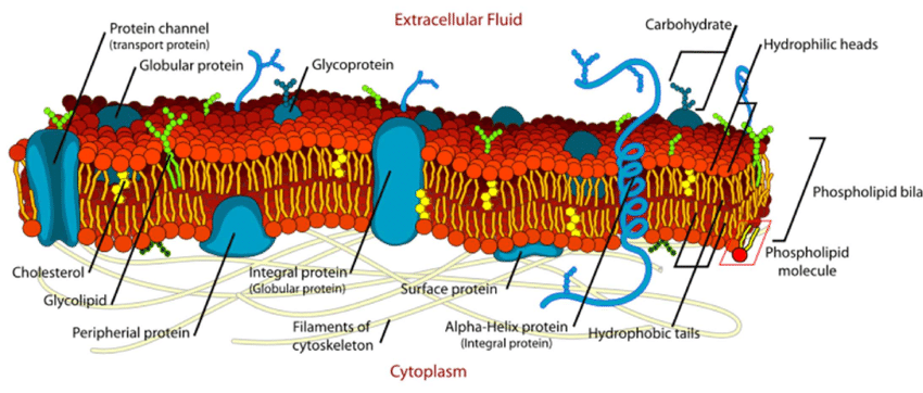
Structure of phospholipid molecule
Polar head
containing negatively charged phosphate group
hydrophilic (water-loving) → interact with water molecule
2 nonpolar fatty acid tails
hydrophobic (water-fearing) → will not mix with water
Hydrophobic tail bury themselves in the center
Hydrophilic heads line up both sides, in contact with water
Components of plasma membrane
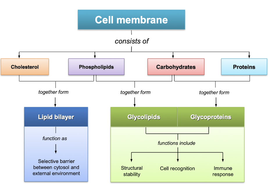
Functions of membrane proteins
structural proteins
transporters
channel proteins
carrier proteins
enzymes
membrane receptor proteins
Functions of plasma membrane
Physical barrier
separate intracellular fluid and extracellular fluid (ECF)
Exchange of materials with the environment
entry of ions and nutrients into cell
elimination of cellular waste and release of products
Communication between cell and environment
surface proteins respond and recognise other molecules
Structural support
cell shape maintained by cytoskeletal proteins attached to membrane proteins
Membrane transport
Different types of membrane transport
Simple diffusion
Movement from high concentration to low concentration
Does not require energy
E.g. gas exchange between cells and ECF in lungs
Osmosis
Diffusion of water molecules across plasma membrane down its concentration gradient
Presence of solutes reduce water concentration
Hypertonic - solution with greater solute concentration
Water travel by osmosis from hypotonic environment to hypertonic environment
Animal cells = crenation (too little water in cells) or hemolysis (too much water, causing cell to burst)
Plant cells = plasmolysis (too little water) or turgid (too much water, but plant cell doesn’t burst because there is cell wall to protect it)
Facilitated diffusion
Similar to simple diffusion, but requires a carrier
Facilitate carrier-mediated transport
Example:
ion channels & aquaporin (water) channels
transport of glucose -- glucose transporter (GLUT)
Active transport
Primary active transport
involves carrier protein
utilises ATP to drive transport of molecules against concentration gradient
Second active transport
utilises potential energy stored in electrochemical gradients of ions to drive transport of another molecule (contransport)
gradients established and maintained by carrier proteins that utilises ATP
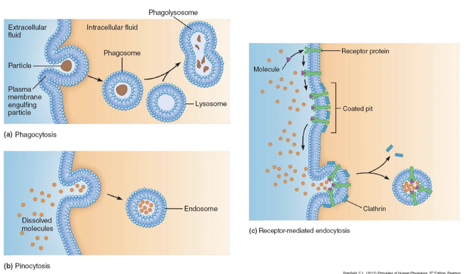
Vesicular transport
Transport of large molecules across membrane
Formation of membrane-enclosed vesicles
Active method of vesicular transport
Endocytosis
Exocytosis
Primary active transport - Na+ - K+ ATPase pump

Cytoplasmic Na binds to pump proteins
Na binding promotes hydrolysis of ATP, energy release during reaction phosphorylates the pump
Phosphorylation causes the pump to change shape, expel Na to the outside
Two extracellular K binds to pump
K binding triggers release of the phosphate, the dephosphorylated pump resumes its original confirmation
Pump protein binds ATP releases K to the inside, and Na site are ready to bind Na again cycle repeats.
Secondary active transport
Co-transport molecules across the plasma membrane
Same direction (symport) - glucose and amino acids
Opposite directions (antiport) - Na and H ions
Endocytosis
Process by which substances move into cell
Phagocytosis - selective uptake of multimolecular particles (e.g. bacteria and cellular debris)
Pinocytosis - nonselective uptake of ECF fluid
Receptor-mediated endocytosis - selective uptake of large molecule (e.g. protein)
Exocytosis
Process by which substance is transported out of cell
Provides mechanism for secreting large polar molecules
Enables cell to add specific components to membrane
Cell-to-cell communication
Physiological signals: Electrical and chemical signals
Basic communications systems:
Direct contact (gap junctions and contact-dependent signals)
Paracrine and autocrine signalling
Endocrine signalling
Synaptic signalling
Direct contact communication
Gap Junctions
Heart, smooth muscle cells, neuron
Protein channels - transfer between adjacent cells
Membrane proteins: Connexins
6 Connexins forms a channel - connexon
Gap junction: Connexons from 2 cells
Contact-dependent signals
Immune cells & Growth/Development
Chemical signalling between cells
Autocrine signal
chemical signals act on cells that secreted it
Paracrine signal
chemicals act on cells in the immediate vicinity of cell secreting the signal
Some molecules may act as both an autocrine and paracrine signal
Signals reach target cell by diffusing through interstitial fluid - distance is the limiting factor for diffusion
Endocrine signals
cells of endocrine glands secrete hormone into ECF, hormones enter blood and carried by blood in cells in body, target cells respond to hormone
Nervous system - combination of chemical and electrical signals to communicate over long distance.
Nerve cells can extend long processes called axons to very near the target cells.
Electric signals travels along neuron until it reaches very end of cell, where it is translated into a chemical signal secreted by neuron.
Neurotransmitter
chemical signals secreted by neurons diffuse accros small gap to target cell and has a rapid effect
Neurohormone
chemicals released by neurons into blood for action at distant targets
Chemical Messengers

Signalling transduction pathway
Describe the mechanisms of signal transduction pathway
lipophilic vs lipophobic messengers
Identify the different functional classes of membrane-bound receptors
Signalling pathway: Receptor protein
Lipophilic (hydrophobic) signal molecules
bind to cytosolic (inside the cell) receptors or nuclear receptors (e.g. transcription in nucleus)
Lipophobic (hydrophilic) signal molecules
stay in the ECF and bind receptor proteins
Membrane-bound receptors
Extracellular signals activates membrane receptors
→ alters intracellular molecules to create cellular response
Three major types:
Channel-linked receptors
similar to sodium-potassium pump, but a pump is about TRANSFERRING materials. A channel-linked receptor has to bind with something first to be able to allow materials to enter/exit.
Change of membrane potential
Acetylcholine, for example, binds to a ligand-gated channel in muscle cells. The binding increases the permeability of the membrane to Na+ and causes it to rush in and depolarize the membrane. The response is brief and does not last very long.
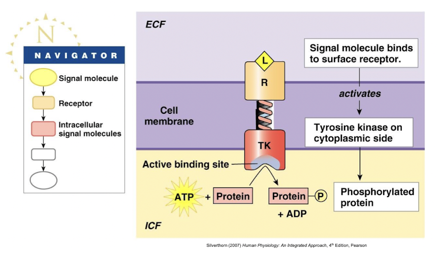
Enzyme linked receptors
Example: Tyrosine kinase
Ligand binds to receptor, which triggers the enzyme, in this case it is tyrosine kinase. Tyrosine kinase adds phosphate onto a particular protein.
G protein-linked receptors
Very specific.
Respond to ligands by activating G proteins
Three subunits: a- (binds to GDP), B- and y subunits
Cytoplasmic tail of receptor linked to G protein
changes from GDP to GTP to become activated
G proteins are activated:
open ion channels in the membrane
alter enzyme activity on cytoplasmic side of membrane - linked to amplifier enzymes
Modulation of signalling pathways
Target cell response - determined by receptor or its associated intracellular pathway
Multiple ligands for one receptor - agonist/antagonist
primary ligand activates a receptor
agonist will also activate the receptor
an antagonist will block receptor activity
Multiple receptor for single ligands
Example: blood vessels
Epinephrine + a-receptor → intestinal blood vessel constricts
Epinephrine + B-receptor → skeletal muscle blood vessel dilates
Termination of signalling pathways
Receptor activity can be stopped in different ways:
Extracellular ligands degraded by enzymes (e.g. protease)
e.g. breakdown of neurotransmitter acetylcholine
Chemical messengers transported back into pre-synaptic cells for recycling
e.g. serotonin re-uptake by transporter protein
Endocytosis of receptor-ligand complex
Ligand removed, receptors returned to membrane as endocytosis (lysis occurs and removes ligand + receptors)
Summary
Describe the structure of plasma membrane with relation to their functions
Identify the different types of membrane transport
Describe the different types of cell-to-cell communication systems
Describe the membrane-bound receptors and mechanisms of signal transduction pathways