Brain Structure and Function The Left and Right Hemispheres The Lobes of the Brain Methods Used to Study the Brain
What are the 3 main parts of the brain?
Hindbrain, midbrain and forebrain
Hindbrain.
Located at the base of the brain, near back of skull. Brain stem.
Responsible for lower-brain functions such as: control of basic autonomic survival function. coordination of voluntary muscle movements.
Main parts include: medulla and cerebellum.
Medulla (oblongata).
Located at the base of the brain stem in front of cerebellum.
Relays information between the spinal cord and brain. Regulates viral involuntary bodily functions.
Cerebellum.
Located at rear of brain stem beneath occipital and temporal lobes.
Helps coordinate voluntary movement, relays motor information.
Midbrain.
Small area in middle of the brain, connects hindbrain and forebrain.
Processes information related to hearing, vision, movement, pain, sleep and arousal. Keeps up alert and attentive.
Main parts include: the reticular formation.
Reticular formation.
No distinct location, spans from lower part of medulla to upper part of midbrain.
Bombards brain with important sensory information, which keeps cerebral cortex active.
Forebrain.
Located above the midbrain towards the top of the brain. Largest most complex and highly developed region of the brain.
Responsible for our most complex processes.
Main parts include: hypothalamus and thalamus.
Hypothalamus.
Located below the thalamus.
Maintains homeostasis. Regulates the release of hormones that help achieve homeostasis. Connects NS to endocrine system.
Thalamus.
Located above hypothalamus, consists of two small egg-shaped structures (thalami), that are found in centre of the brain.
Relay system for sensory messages going towards the cerebral cortex. Conducts motor signals.
Define cerebrum.
Lies over and around most brain structures. It is responsible for our most complex processes.
Define cerebral cortex.
The outer layer, or surface, of the cerebrum.
What is the left hemisphere responsible for?
Controls the functions of the right side of the body and is responsible for the verbal and analytical functions.
Language function.
The ability to communicate our awareness using spoken or written language depends on left hemisphere function.
Analytical function.
Breaks information into parts and processes is sequentially. Focuses on small details. Involved in a person having superior ability to math, judging time and rhythm and coordinating movements.
What is the right hemisphere responsible for?
Controls the functions of the left side of the body and is responsible for the non-verbal functions, which are:
information processing
non-verbal communication
spatial skills
Information processing.
Processes information simultaneously and holistically. Involved in a person having superior ability at assembling pieces of information.
Non-verbal communication.
More dominant in detecting emotion non-verbally. Contributes to the understanding of language.
Spatial skills.
Right hemisphere is dominant in spatial and visual skills such as recognising patterns and faces. Creative side of the brain.
Contralateral control of the body.
How each side (hemisphere) of the brain controls the opposing side of the body.
What is the corpus callosum?
The thick bundle of nerve fibres that connect your left and right hemispheres.
Found deep inside the centre of the brain and ensures both sides of the brain can communicate with each other.
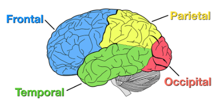
What are the 4 lobes of the brain?
Frontal, parietal, occipital, temporal.
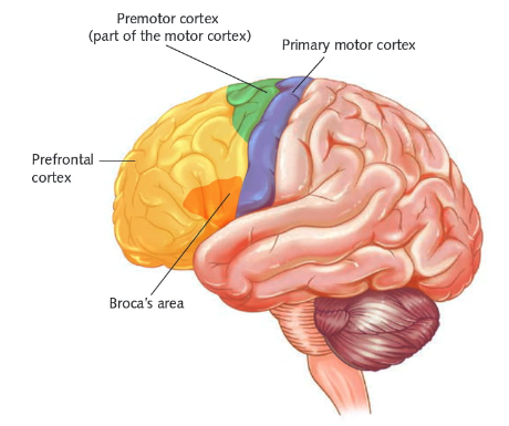
Parts of the frontal lobe.
Pre-frontal cortex, primary motor cortex, broca’s area (only in the left frontal lobe).
What’s the role of the pre-frontal cortex?
Higher level cognitive functions (planning, decision making, problem solving etc.)
Predicts possible consequences of actions using memory to control responses.
Work with the amyglala and hippocampus to regulate emotions.
What’s the role of the primary motor cortex?
Controls voluntary movement of skeletal muscles.
Left PMC controls right side of body.
Right PMC controls left side of body.
What’s the role of the broca’s area?
Controls production of articulate speech.
Controlling the muscles in jaw, cheeks, lips, tongue and even diaphragm that produce the words.
Damage = broca’s aphasia (unable to produce clear and articulate speech).
Parts of the parietal lobe.
Primary sensory cortex.
What’s the role of the primary sensory cortex?
Also called somatosensory cortex.
Processes and register’s sensations detected by sensory receptors.
A strip of neurons located at the front of the parietal lobe.
Parts of the occipital lobe.
Primary visual cortex.
What’s the role of the primary visual cortex?
Registers and processes visual information transmitted from the retinas of your eyes.
Specialised neurons in association areas select and integrate information from the primary visual cortex, and send visual information to other brain lobes.
Parts of the temporal lobe.
Primary auditory cortex, wernicke’s area.
What’s the role of the primary auditory cortex?
Registers and processes auditory information received by the ears.
Primary auditory cortex in the right temporal lobe specialises in processing non-verbal sounds.
Primary auditory cortex in the left temporal lobe specialises in processing verbal sounds.
What’s the role of wernicke’s area?
It identifies sounds as words so their meaning can be understood.
It accesses words stored in memory so it controls comprehension of speech and the formulation of meaningful sentences.
What are the 4 methods to study the brain?
Electroencephalogram, computer tomography, magnetic resonance imaging and functional magnetic resonance imaging.
Describe electroencephalogram (EEG).
Technique involving electrical activity. Measures the electrical activity of the brain (brain waves) using small metal discs (electrodes) that are attached to the scalp.
Describe computer tomography (CT).
Technique involving radiation. CT scans are x-rays of the brain that produce an image of the brain structure.
Describe magnetic resonance imaging (MRI).
Technique involving magnetic fields. MRI scans use strong magnetic fields and radio waves to produce a detailed image of the brain structure.
Describe functional magnetic resonance imaging (fMRI).
Technique involving magnetic fields. A specialised form of MRI. Used to examine the brain’s functional anatomy, meaning the part of the brain that handles critical functions.