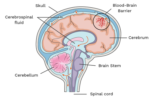
How is the brain protected and what are the functions of meninges, CSF, and blood brain barrier (BBB)?
Skull
Meninges = membranes; connective tissue membranes, external to CNS organs
Functions:
Cover & protect CNS
Protect blood vessels and enclose venous sinuses
Contains CSF
Form partitions in the skull
Cerebrospinal fluid = water cushion
clear fluid
found in around the brain and spinal cord & protects from trauma and blows
Forms a liquid cushion around the CNS structures
Prevents the brain from crushing under its own weight
Carries nutrients
Blood-brain barrier = protects the brain from harmful substances
Brain is reliant on a constant internal environment
Consists of tight junctions between capillary endothelial cells
Functions:
Maintains homeostasis in brain
Selectively filters wanted & unwanted nutrients between the brain’s capillaries and the brain
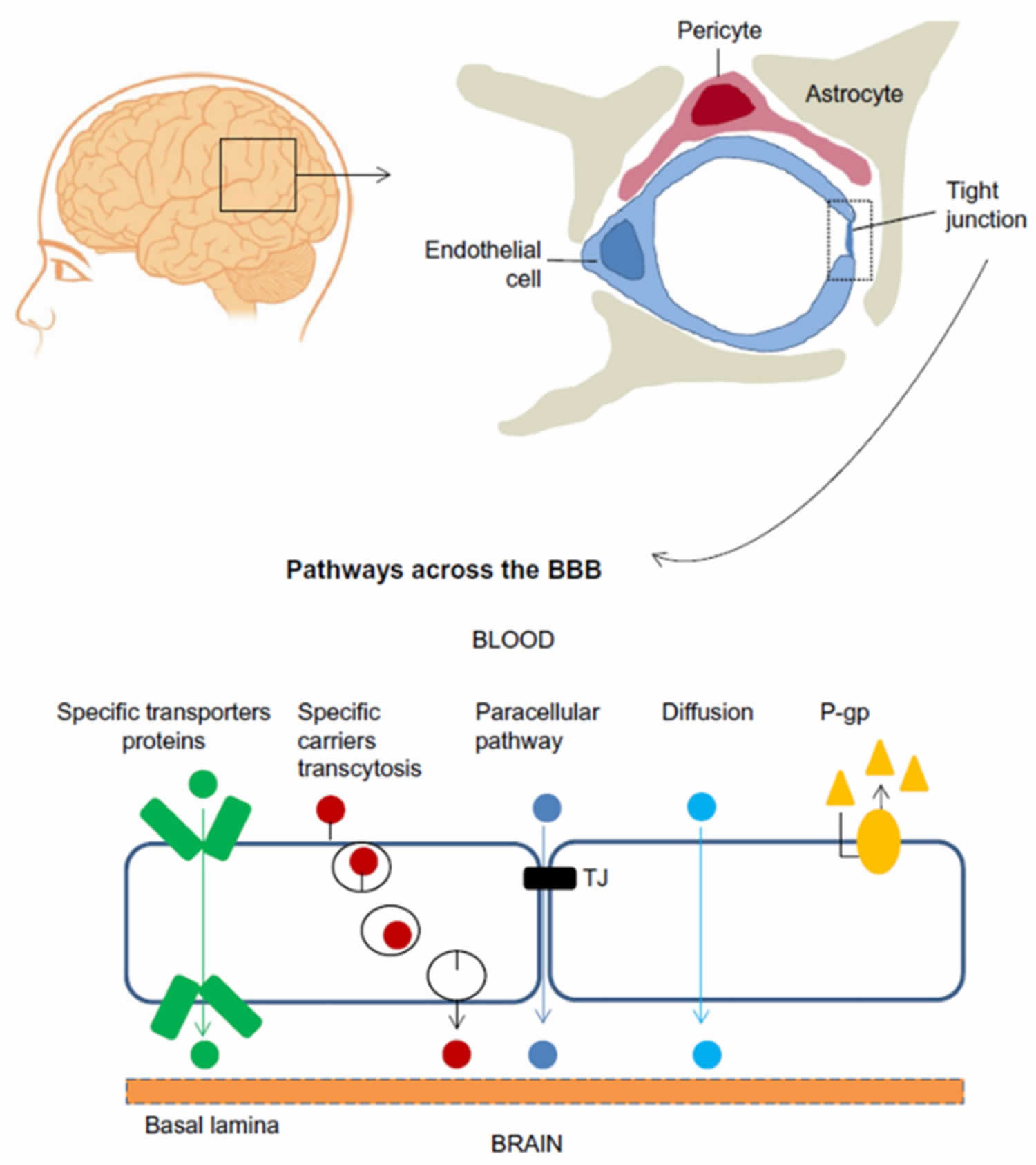
What neuroglia assist the Blood Brain Barrier and how does BBB work (fig 12.26).
Astrocytes cling to each capillary and provide structural support for neurons.
BBB - is the protective mechanism that helps maintain the brain’s stable environment
Impermeable tight junctions between capillary endothelial cells are its major component
Pericytes - surrounding the endothelial cells are the bulbous feet of astrocytes and smooth muscle
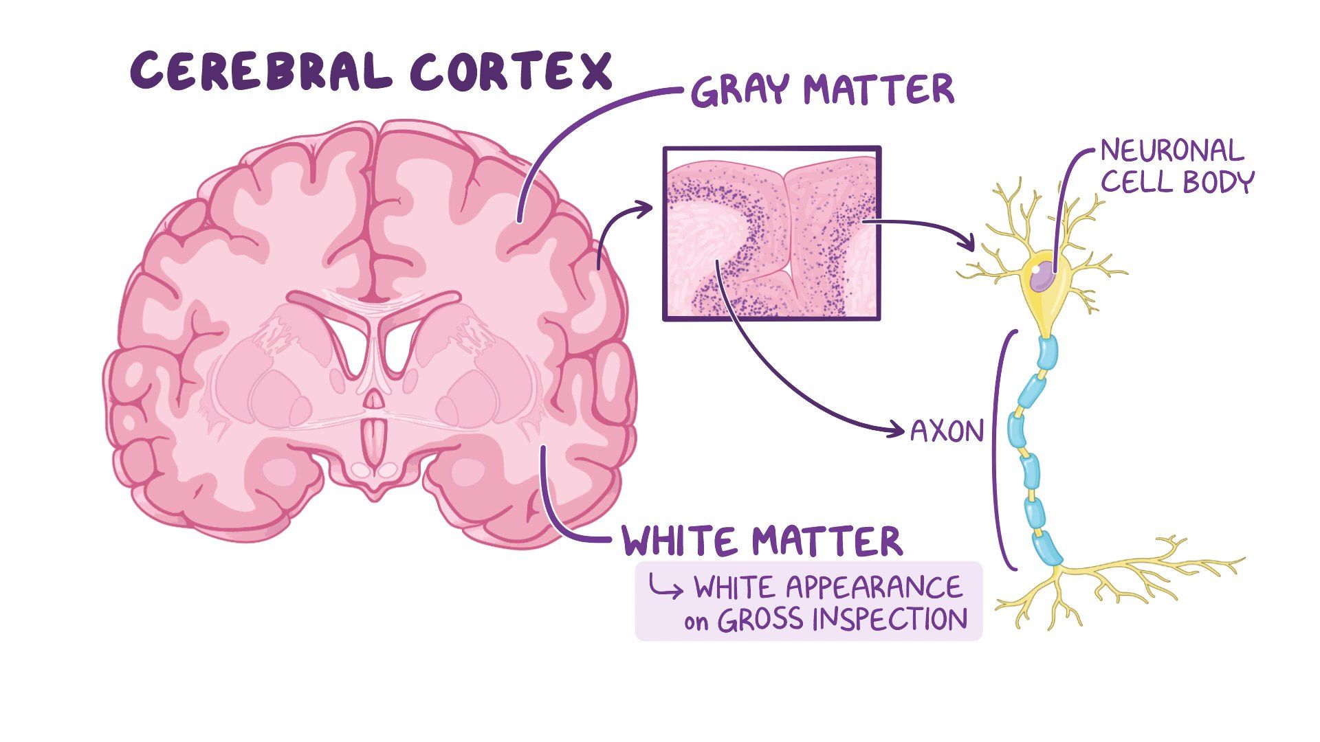
What is white and gray brain matter?
White - consists mostly of myelinated axons with some nonmyelinated axons primarily in fiber tracts. The dense coating of fatty myelin is what gives white matter its color
Gray - consists of short, nonmyelinated neurons & neuron cell bodies
The spinal cord exhibits this basic pattern. Charges with ascent into the brain stem
The brain stem has additional gray matter nuclei scattered within the white matter
The cerebral hemispheres and the cerebellum have an outer layer or “bark'“of gray matter called a cortex
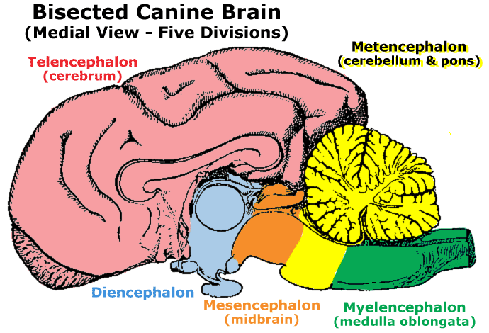
What are five major divisions of the brain?
Telencephalon - the largest division of the brain
Contains the cerebrum, which constitutes 2/3 of the brain’s mass
Also includes the olfactory and optic cranial nerves
Diencephalon - relays sensory information and connects the endocrine system with the nervous system
Regulates functions such as autonomic control, endocrine signaling, and sensory perception
Mesencephalon (midbrain)
Sits between the forebrain & hindbrain
Essential for visual and auditory processing
Involved in reflexive movements, such as turning toward a sudden sound
Metencephalon (afterbrain)
Contains the pons & the cerebellum
Pons: Connects different brain regions & help regulate breathing
Cerebellum: Coordinates motor movements, balance, and posture
Myelencephalon (hindbrain) (spinal brain)
Includes the medulla oblongata
Vital for autonomic functions like heart rate, breathing, and blood pressure regulation
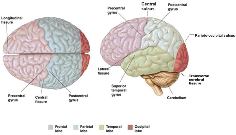
What are gyri, sulci and fissures?
Gyri (plural)
twisters
Elevated ridges of tissue
Sulci (plural)
furrows
shallow grooves
Fissures
Deeper grooves
Separate deeper regions of the brain
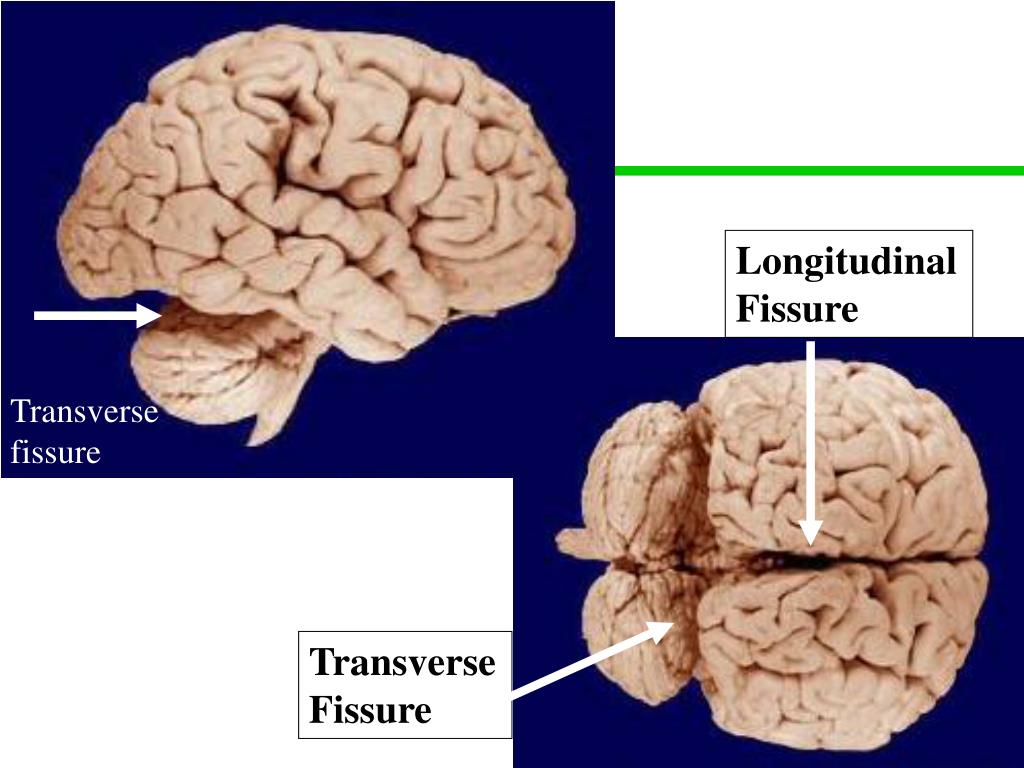
What are longitudinal and transverse fissures?
Longitudinal fissure - separates left & right hemispheres
Transverse cerebral fissure - separates cerebral hemispheres from cerebellum
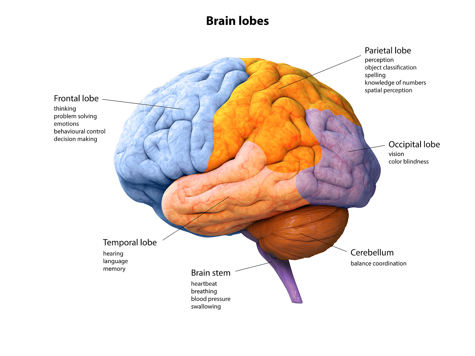
What five lobes of cerebrum and what is each major functions?
Frontal - smell, voluntary motor function, motivation, aggression, and mood
Parietal - Receive general sensory input, taste, and balance
Occipital - visual centers
Temporal - receive olfactory & auditory input, involved with memory, abstract thought, and judgement
Insula - deep to portions of the temporal, parietal, and frontal lobes
island - located deep within the tissue separating the temporal lobes from the others
Most lobes are named for the cranial ones that cover them
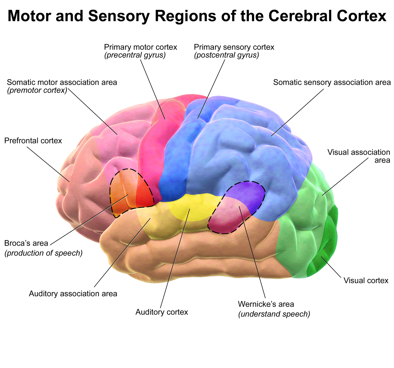
What/where is the cerebral cortex, and how is it organized/structured?
is the executive suite of the nervous system, where our conscious mind is found
Composed of gray matter
“Higher order” functions
Self-awareness, communication, memory, understanding, voluntary movements
3 functional areas:
Motor
Sensory
Association
What does each of the three subsections of the cortex do?
White matter
*Mostly myelinated axons with some nonmyelinated axons
Basal nuclei = clusters of cell bodies
Neuron cell bodies
Short nonmyelinated neurons
Ventricles – filled with CSF, lined with ependymal cells nonmyelinated
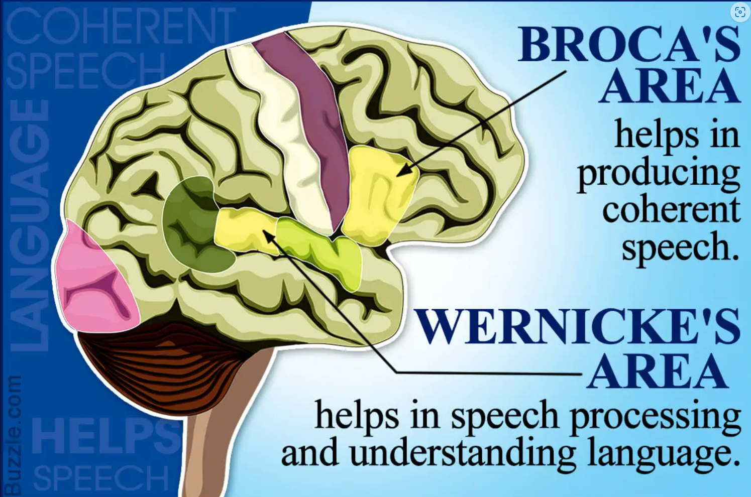
What are Boca’s and Wernicke’s areas of brain?
Boca’s Area
Usually only present in left hemisphere
Special “motor speech area”
Wernicke’s Area
Speech word bank
Direct muscles involved with speech production
Active when we prepared to speak and even as we think about (plan) many voluntary motor activities other than speech
What is the motor somatotopy and why is this important?
Somatotopy - spatial representation & mapping of the entire body
Homunculus - is used to show the broad areas of the primary cortex
Most precise motor control - face, tongues, & hands & consequently those areas are the largest, represented by their size on the homunculus
What is the somatosensory cortex of brain and how is it organized?
Integrates & creates an understanding of sensory input
Temperature, pressure so forth from the primary somatosensory cortex to produce an understanding of an object being felt-size texture, etc
Understanding is based on learned/past experiences
Know without seeing; know what we feel
List and define the 12 Sensory areas of the Cortex?
1) Primary visual cortex (V1) - located in the occipital lobe, responsible for processing visual information
largest areas of all retinal images, focal images
2) Secondary visual cortex (V2, V3, V4, V5/MT)
located in the occipital lobe, responsible for higher-level visual processing
3) Primary auditory cortex - located in the temporal lobe, responsible for processing auditory information
pitch, loudness, location in 3D space
4) Somatosensory cortex - located in the frontal lobe
integrates senses of temperature, touch, pressure, size, and texture, (know without seeing; know what we feel)
5) Primary motor cortex - located in the frontal lobe, responsible for controlling voluntary movements
precise, skilled motions
6) Premotor cortex - located in the frontal lobe, involved in planning and coordinating complex movements
learned, repetitive motor skills
7) Supplementary motor area - located in the frontal lobe, involved in the preparation and initiation of voluntary movements
8) Gustatory cortex - located in the parietal lobe, responsible for processing taste information
perception of taste, located in the insula
9) Olfactory cortex -
sense of smell
10) Vestibular cortex - Equilibrium; a position sense of where your head is in space
located in the parietal lobe, responsible for processing information about balance and spatial orientation
11) Insular cortex - located in the temporal lobe, involved in processing interoceptive
12) Posterior parietal cortex - located in the parietal lobe, involved in integrating sensory information and spatial awareness
What occurs in the multimodal, anterior, posterior, limbic areas of cerebrum?
Multimodal - allows us to give meaning and content to what is experienced; where thoughts and emotions become “real” to us; and we store it in memory. It is our collective personality.
a. Anterior Association area (prefrontal cortex) – intellect, complex reasoning, ideas, judgement, planning
b. Posterior Association Area – recognizing patterns & shapes, where we are in the big “picture”
c. Limbic Association area – provides the emotional impact of events to us
Define lateralization, contra lateralization and cerebral dominance?
Lateralization - we use both cerebral hemispheres for almost every activity, and the hemispheres appear nearly identical.
Nonetheless, there is a division of labor, and each hemisphere has abilities not completely shared by its partner.
Contra laterilization - property that the hemispheres of the cerebrum and the thalamus represent mainly the contralateral side of the body
Cerebral hemisphere - "dominates" each task and designates the hemisphere that is dominant for language.
In most people (about 90% ), the left hemisphere has greater control over language abilities, math, and logic
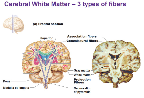
What do the ‘Fibers” of the cerebral white matter do?
Located deep to cortical gray layer
Responsible for communication between cortex and lower CNS centers
Consists of myelinated fiber tracts:
Commissures: connect corresponding gray areas of different hemispheres
Association fibers: connect different parts of the same hemisphere
Projection fibers: tie the cortex to the rest of the nervous system/body
What does basal ganglia do?
Basal Ganglia
Located deep within white matter
Starting, stopping, and regulating the intensity of movements executed by the cortex
Act as “filters” for incorrect/inappropriate responses
Disorders of the basal nuclei may result in increases or decreased movement
i.e. Parkinson’s Disease
What structures comprise the diencephalon and what do they govern?
Located between the brainstem and the cerebrum
Thalamus
Greek = “inner room”
Acts as a relay center for information between the cortex and the rest of the body
Edits and sorts out information
Mediates sensory, motor, and cognitive functions
“the gateway to the cortex”
Hypothalamus
Visceral control center
Regulation of homeostasis
Heart rate, blood pressure, digestive function, pupils (ANS)
Regulates emotion in conjunction with limbic system
Sleep wake-cycles
Body temperature
Located ‘below’ the thalamus
Vitally important to homeostasis
Epithalamus
Pineal gland
Secretes melatonin
Assists hypothalamus with sleep-wake cycle
Melatonin is a hormone that serves as a sleep-inducing signal and antioxidant
What structures comprise the brain stem and what are their functions?
Midbrain
Conduction pathway between higher and lower brain centers
Cerebral peduncles
Major descending motor pathway
Lies between the diencephalon and the pons
Pons
Conduction pathway between higher and lower center
Regulates:
Respiration
Communication/connects between cerebrum and cerebellum
Medulla oblongata
Sensory impulses from skin & proprioceptors
Heart rate
Vessel diameter
Respiratory rate
Hypothalamus and Medulla have a lot of overlap. Hypothalamus controls the functions by relaying the messages to the medulla oblongata which carry it out.
CV Center- adjusts the force and rate of heart contraction, vasodilation
Respiratory rhythm, rate, and depth
What is function of the cerebellum?
Coordination between gross and fine motor tasks
Provide instructions to motor cortex
Balance
Capable of learning complex motor activities
Provides instructions to cerebral motor cortex, resulting in smooth coordinated skeletal muscle movements
How does cerebellum processing work?
1. Cerebellum receives impulses (notifications of “intent” from cortex)
2. Proprioceptors & visual signals inform cerebellum of body’s positioning
3. Cerebellum decides proper movement and calculates the best way to coordinate the force, direction, and extent of the muscle contraction
4. Sends plan (blueprint) to cerebral motor area for execution
What is the limbic system and what does it do?
is a group of structures located on the medial aspect of each cerebral hemisphere and diencephalon. Its cerebral structures encircle (limbus=ring) the upper part of the brain stem.
Functions:
Emotion Regulation: it helps regulate emotions and behaviors, managing how we feel and respond to certain situations or stimuli
Behaviors: They influence our behavior and motivation, often in response to emotional stimuli
Memory: It plays a key role in forming new memories, particularly those tied to emotions
Autonomic Nervous System: it interacts with the autonomic nervous system, affecting physiological responses like heart rate, blood pressure, & digestion
Higher Mental Functions: the limbic system works with other brain regions to support higher mental functions like learning, speech, and decision-making
Involved in..
Hippocampus - involved in memory
formation and spatial navigation
Amygdala - responsible for emotional responses, fear, and aggression
Thalamus - relays sensory information from the top down to the correct brain regions
Hypothalamus - regulates hunger, thirst, body temperature, and other autonomic functions
Cingulate gyrus - associated with emotional processing & decision-making
What is reticular formation and what does it do?
is a neuron network in the brainstem that enables communication, sensory, & motor function, and endocrine & neurotransmitter regulation
Includes 2 main systems:
(ARAS) - Ascending Reticular Activating System - This system sends sensory signals to the cortex, affecting alertness and wakefulness.
(DRS) - Descending Reticular System - this sytem sends signals to the motor neurons of the spinal cord & can influence muscle tone & reflexes
What are declarative memory, procedural memory, emotional memory and motor memory?
Declarative memory - (fact) (explicit memory) names faces, words, and dates
Procedural memory - (skills) (piano playing)
Motor memory - riding a bike (motor skills)
Emotional memory - pounding heart/emotional memories are closely tied to our feelings & emotions
What influences our ability to move topics from short term to long term memory?
Short-term memory (STM) - working memory is the preliminary step of looking at a phone number and never thinking about it again
Long-term memory (LTM) - seems to have limitless capacity. Long-term can be forgotten, and so our memory bank continually changes over time.
What influences our ability is…
Emotional state: We learn best when we are alert, motivated, surprised, or aroused
Rehearsal - rehearsing or repeating the material enhances memory
Association- tying “new” info to “old” information already stored in LTM appears to be important in remembering formed
Automatic memory - not all impressions that become part of LTM are consciously formed
What is consciousness? How is EEG related to consciousness in terms of alpha, beta, theta, delta waves?
encompasses perception of sensations, voluntary initiation and control of movement, and capabilities associated with higher mental processing (memory, logic, judgement, and so on).
Brainwaves are electrical oscillations produced by the synchronized activity of neurons in the brain.
EEG measures the frequency (cycles per second or hertz) and amplitude (strength) or brainwaves.
Alpha - awake & resting (8-13 Hz)
Beta - mental activity (14-30 Hz)
Theta - emotional stress (4-7 Hz)
Delta - deep sleep (4 Hz or less)
How is sleep regulated? What is the importance of sleep?
2 major types of sleep:
non-REM (NREM) - NREM sleep is essential for physical restoration, tissue repair, and immune system function.
REM (rapid eye movement) - REM sleep is crucial for cognitive processes, memory consolidation, and emotional regulation.
Circadian Rhythm: Sleep is regulated by our circadian rhythm, which is like an internal clock that influences when we feel sleepy or alert.
The importance of sleep is …
During sleep, we may consolidate new memories and discard memories that are no longer accessed (in other words, we forget).
Sleep is presumed to be a restorative time when most neural activity can wind down to basal levels.
When deprived of sleep, we spend more time than usual in both REM and slow-wave sleep during the next sleep episode in an attempt to catch up.
Define the cross-sectional anatomy of the spinal cord and its anterior/posterior spinal horns?
The spinal cord is somewhat flattened from front to back & two grooves mark its surface: the wide ventral (anterior) median fissure and the narrower dorsal (posterior) median sulcus
Where does CNS end and PNS begin within the spinal column?
CNS and the PNS within the spinal column is at the point where the spinal nerves exit the spinal cord.
The spinal cord itself ends at about the first or second lumbar vertebra (L1-L2) in adults, but the nerves continue to extend down the vertebral column forming the cauda equina.
The CNS ends and the PNS begins at the intervertebral foramina, where the nerve roots exit the spinal cord and extend out to the body
How is gray matter in spinal cord organized?
Connects brain and peripheral nervous system (PNS)
Enclosed in the vertebral column
Two-way conduction pathway to and from the brain
Gray matter
“H” or “butterfly” shape
Dorsal projections = dorsal (posterior) horns
Ventral projections = ventral (anterior) horns
Forms columns; running the entire length of the cord
Generally
Dorsal horns = sensory function
Ventral horns = motor function
Dorsal root & dorsal ganglion (enlarged area)
Serve as sensory afferent
Ventral root and ventral ganglion
Serve as motor efferent
What is paraplegia, quadriplegia?
Paraplegia - transection of the spinal cord between T1 and L1 (Thoracic vertebra)
- paralysis of the legs and lower body, typically caused by spinal injury or disease.
Quadriplegia - transection of spinal cord in the cervical region
paralysis of all four limbs
What is decussation, relay, somatotopy?
Decussation - Most pathways cross from one side of the CNS to the other (decussate) at some point along their journey
Relay - most pathways consist of a chain of two or three neurons (a relay) that contribute to successive tracts of the pathway
Somatotopy - most pathways exhibit somatotopy, a precise spatial relationship among the tract fibers that reflects the orderly mapping of the body
What do 1st, 2nd and 3rd order neurons do in the ascending pathway?
First-order neurons
Impulses from cutaneous receptors and proprioceptors
dorsal root ganglion
Second-order neurons
Form dorsal horn —> thalamus
Third-order neurons
Relay impulses from thalamus to somatosensory cortex
In general what do the “tracks” in the spinal cord represent (12.3 table)?
These tracts carry sensory information & motor stimuli from the periphery (body) to the brain.
CHP 13
Know the classification of receptors by stimulus type and location; this includes mechanoreceptors, thermoreceptors, photoreceptors, chemoreceptors, nociceptors, exteroceptors, interoceptors., proprioceptors
Mechanoreceptors - respond to touch, pressure, vibration, stretch, tension, and itch
Thermoreceptors - temperature
Photoreceptors - photo
Chemoreceptors - chemicals
Nociceptors - pain
Exteroceptors - sensitive to stimuli arising outside the body
touch, pressure, pain, temperature receptors of skin & most receptors of sense organs
Interceptors - respond to stimuli arising within the body
chemical changes, stretching of tissues & temperature
Proprioceptors - Respond to internal stimuli expect their location is restricted to musculoskeletal organs
Provide the brain information to location of body in space (kinesthetic awareness)
Know the location and functions of: tactile receptors, muscle spindles, tendon organs, joint kinesthesia receptors
Tactile receptors -
Functions: mechanoreceptors
Location: exteroceptors; fingertips, feet, and eyelids
Muscle spindles -
Functions: mechanoreceptors; muscle stretch, and length
Location: proprioceptors; skeletal muscles
Tendon organs
Functions: Mechanoreceptors (tendon, stretch, tension
Location: proprioceptors, tendons
Joint kinesthesia receptors -
Functions: Mechanoreceptors & nociceptors
Location: joint capsules of synovial joints
What is the somatosensory system and its functions?
A part of the sensory system serving the body wall and limbs — receives inputs from exteroceptors, proprioceptors and interceptors
Functions:
Touch - this include the perception of pressure, vibration, and texture
Temperature - the system detects variations in temperature (thermoception)
Pain - it processes nociceptive signals which are essential for avoiding injury
What are the three main levels of the somatosensory system and how does each work.
Receptor level: sensory receptors
For sensation to occur, a stimulus must excite a receptor, and action potential must reach the CNS
Circuit level: processing in ascending pathways
the task is to deliver impulses to the appropriate region of the cerebral cortex for localization and perception of the stimulus
Perceptual level: processing in cortical sensory areas
the ability to identify and appreciate sensations depends on the location of the target neurons in the sensory cortex, not on the nature of the message
What is encoding/transduction?
Encoding - refers to the initial step in creating a memory. It involves transforming sensory input from the environment into a form that can be stored in the memory system
Transduction - describes the conversion of physical signals from the environment into neural signals that are sent to the CNS
What is neuronal adaptation?
Gradual decrease over time in the responsiveness of the sensory system to a constant stimulus
What is perceptual detection?
Perceptual detection - is the ability to detect that a stimulus has occurred. This is the simplest level of perception. As a general rule, inputs from several receptors must be summed for perceptual detection to occur.
What is magnitude estimation?
Magnitude estimation - is the ability to detect how intense the stimulus is. Perceived intensity increases as stimulus intensity increases because of frequency coding
What is spatial discrimination?
Spatial discrimination - allows us to identify the site or pattern of stimulation
What is feature abstraction?
Feature abstraction - is the mechanism by which a neuron or circuit is tuned to one feature, or property of a stimulus in preference to others
What is quality discrimination?
is the ability to differentiate the sub modalities of a particular sensation
What is pattern recognition?
is the ability to take in the scene around us & recognize a familiar pattern, an unfamiliar one, or one that has special significance for us
What roles do substances p, histamine, glutamate, endorphins, and enkephalins have in pain sensations?
Substance p - is a neuropeptide involved in regulating nociception (pain sensation) in the body
it acts as a sensory neurotransmitter in the spinal cord interneurons, resulting in the suppression of pain
Histamine - is a chemical mediator released during allergic reactions & inflammation
it plays a role in pain by sensitizing nerve endings and increasing their responsiveness to other pain signals
Glutamate - is a major excitatory neurotransmitter in the CNS
Endorphins - are endogenous opioids produced by the body
they act as natural painkillers by binding to opioid receptors in the brain and spinal cord
Enkephalins - are another group of endogenous opioids
bind to opioid receptors & modulate pain signals
What are the 12 cranial nerves, their common name, their roman numeral and their major functions?
Olfactory (I) nerve
Smell
Surely sensory
Optic (II) nerve
Vision
Purely sensory, carry afferent impulses for vision
Oculomotor (III) nerve
Innervates four of six eye muscles and the upper eyelid
Eye movement & pupillary reflex
Mostly motor - movement of the eye
Parasympathetic - ANS motor fibers to pupils which caus pupls to constrict
Test of this nerve shine penlight into eye pupils shouldd constrict when illuminated
Trochlear (IV) nerve
Eye movement
Primary motor nerves; one extrinsic eye muscle
Trigeminal (V) nerve
Muscles of mastication
Has greatest cutaneous sensory distrubution of any cranial nerve
Sensory impulses of anterior scalp, eyelids, nose, cheek, teeth
3 branches, 2 of which innervate the teeth
Motor - chewing muscles
Novocain - largest cranial nerve; used to block pain transmitting fibers of the teeth. Surrounding tissues becomes numb
Abducens (VI)
Extrinsic eye muscle
Turns the eyeball laterally (abducts)
Abduction of the eye
Facial (VII) nerve
Supplies muscles of facial expression
Involved in sense of taste
Salivary glands and lacrimal glands
Taste, facial expression, tearing
Tearing is controlled by the ANS
Bell’s palsy - sudden onset characterized by inflamed or swollen faciial nerve
Symptoms - paralysis or facial muscles on affected side
Vestibulocohelar (VIII) nerve
Involved in hearing & balance
Mostly sensory
Vestiular branch transmits afferent impulses for sense of equilbrium
Cochlear branch transmits afferent impulses for sense of hearing
Small motor component adjusts the sensitivity of the senroy receptors
Glossopharyngeal (IX) nerve
Swallowing, gag reflex
Sensory info from the:
Posterior tongue (taste)
Pharnyx = muscular tue extending from nasal cavity to esophagus
Vagas (X) nerve
Innervates muscles of the pharnyx and larnyx (voice box)
Swalloing and speech
Fibers supply heart, lungs, abdominal viscera- only cranial nerve wih visceral connections
ANS regulation of HR, breathing, digestion
Accessory (XI) nerve
Thought to have cranial that helped (accessory) the Vagas nerve
Not the case
Trapezius - shoulder elevation (shoulder shrug test is often used to test the health of the accessory nerve)
Hypoglossal (XII) nerve
Motor function - tongue muscles
Allow tongue movements that mix and manipulate food during chewing
What is the role of nerve plexus in the body?
the ganglia interconnected by unmyelinated fiber tracts sorted and travels to anatomical structures to communicate signals to and from your brain
receives more than 1 spinal nerve
What are dermatomes?
is an area of skin innervated by the cutaneous branches of a single spinal nerve
What are the components of a reflex arc and what does each do?
Receptor - site of stimulus
Sensory neuron - transmits the afferent neurons to the CNS
Integration center - either monosynaptic of polysynaptic region without the CNS
Motor neuron - conducts efferent impulses from the integration center to an effector
Effector - muscle fiber or gland that responds to the efferent impulses
Monosynaptic - single synapse between sensory and motor neuron
Polysynaptic - multiple synpases, with chains of interneurons
List the functions of the following reflexes: stretch reflex, tendon reflex, crossed extensor reflex
Stretch Reflex - muscle spindles
Proprioceptors that communicate info about length of a muscle
Rapid forceful stretching activates the spindle
Causes contraction when resists the stretching
Tendon Reflex - muscle relax and lengthen Golgi tendon organs
provide information about the amount of tension in muscle
Contracting the muscle activates the Golgi tendon organs
Afferent Golgi: tendon neurons are stimulated, neurons inhibit the contracting muscle
As a result, the contracting muscle relaxes and the antagonist contracts
Crossed Extensor Reflex - two parts
The stimulated side is withdrawn (away) Flexor Reflex
The opposite side is extended
Flexor Reflex - can be overridden by higher brain centers, such as when you are expecting a painful stimulus (finger stick) for example to draw blood)
Includes initial flexor reflex followed by crossed extensor on the opposite of the body
CH 14
__________________________
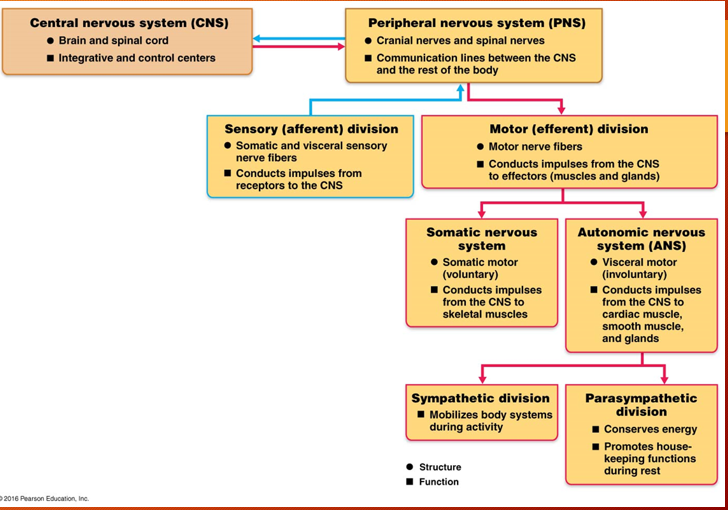
Draw the structural organization of the CNS and PNS and its subdivisions. Know how these subdivisions control behaviors.
The picture >
Figure 14.2 represents a summary of the parasympathetic and sympathetic system and shows how the nerves are organized, their connections and their neurotransmitter; know how and why these connections occur and how this benefits each system as compared to the somatic system.
The picture
What is the role of the sympathetic system?
ganglion located close to CNS and away from target organs = short preganglionic fibers & long post-ganglionic fibers
Mobilizes the body activity
E division
Exercise, embarrassment, excitement, emergency
Vigorous physical activity
Blood is directed towards active skeletal muscles
Visceral blood vessels are constricted
What is role of parasympathetic system?
ganglion located away from CNS and near target organ = long preganglionic fibers
Promotes maintenance functions and conserves body energy
Reason its important to rest and not exercise after a big meal
D division
Digestion, Defecation, Diuresis (urination)
Directs general “housekeeping” activities
For each ANS system, know the following: (table 14.1 and fig 14.3, 14.4, 14.5): What are the sites of origin of the fibers , relative length of fibers, location of ganglia
SITES OF ORGANS
PARASYMPATHETIC
Emerge from the brain (cranial nerves)
Ganglion located away from CNS and near target organ = long preganglionic fibers, and short pre-ganglionic fibers
SYMPATHETIC
Originate in the thoroculumbar region of the spinal cord
Ganglion located close to CNS and away from target organs = short pre-ganglionic fibers and long post-ganglionic fibers
This functional arrangements means -sympathetic system can take a signal and initiate a “all hands-on deck” command and gets a very rapid, collective response.
Preganglinoic cell bodies arise from thorasic and first 2 lumbar segments of spinal cord (T1-T2)
RELATIVE LENGTHS OF FIBERS
PARASYMPATHETIC
Long pre-ganglionic and sort post-ganglionic fibers
SYMPATHETIC
The opposite
Short preganglinoic fiber and long post ganglionic fibers
LOCATION OF GANGLIA
PARASYMPATHETIC
Located in the visceral
Preganglinoic neuron long synapses with post ganglinoic neuron near effector
SYMPATHETIC
Lie close to the spinal cord
Preganglinoic neurons are short. Synapses with post ganglinoic neuron near spinal cord
What are the two types of Cholinergic receptors; what happens when ACh binds to each one of these?
Cholinergic receptors - release acetylcholine from preganglionic neurons & parasympathetic post-ganglionic neurons
Nicotinic - receptors response to ACh binding is always stimulatory
Muscarinic - receptors response to ACh binding is either stimulatory or inhibitory depending on the subclass of muscarinic receptors on the target organ
Nicotinic & Musacarnic are named for drugs that bind to them and mimic acetylcholine’s effects
What are Adrenergic receptors? What happens when NE binds to these receptors?
Adrenergic neurons release norepinephrine (NE)
From postganglionic sympathetic neurons only
Excites or inhibits organs depending on receptors
Organs that respond to NE have one or more of the following receptors:
Alpha I and Beta I receptors produce excitation
Alpha 2 and Beta 2 receptors cause inhibition
What does Parasympathetic and Sympathetic divisions have on the following: eye iris, sweat glands, arrector pilli muscles, heart muscles, lungs, digestive system, blood vessels, cell metabolism, adipose tissues.
Eyes (Iris)
PARASYMPATHETIC
Stimulates sphincter pupillae muscles; constricts pupils
SYMPATHETIC
Stimulates dilator pupillae muscles; dilates pupils
Sweat Glands
PARASYMETHETIC
No effect (no innervation)
SYMPATHETIC
Stimulates copious sweating (cholinergic fibers)
Arrector pilli muscles attached to hair follicles
PARASYMPATHETIC
No effect (no innervation)
SYMPATHETIC
Stimulates contraction (erects hairs and produces “goosebumps”
Heart Muscles
PARASYMPATHETIC
Decreases rate (slow heart)
SYMPATHETIC
Increases rate and force of heartbeat
Lungs
PARASYMPATHETIC
Constricts bronchioles
SYMPATHETIC
Dilates bronchioles
Digestive System
PARASYMPATHETIC
Increases motlity (perstalsis) and amount of secretion by digestive organs; relaxes sphincters to allow food to move through tracts
SYMPATHETIC
Decreases activity of glands & muscles of the digestive system; constricts sphincters; anal sphincters
Blood Vessels
PARASYMPATHETIC
Little or no effect (expect blood vessels to external genitalia)
SYMPATHETIC
Constricts most vessels & increases blood pressure; constricts vessels of abdominal viscera and skin to divert blood to muscles, brain, and heart when neccesary
Cell Metabolism
PARASYMPATHETIC
No effect (no innervation)
SYMPATHETIC
Increases metabolic rate
Adipose tissue
PARASYMPATHETIC
No effect (no inntervation)
SYMPATHETIC
Stimulates lipolysis (fat breakdown)
What is vasomotor tone; what is parasympathetic tone and how do these work together with regard to heart and blood flow.
Vasomotor Tone - the baseline level of constriction or dilation in blood vessels
Function: It helps maintain vascular resistance and blood pressure
The vasomotor raises your heart rate (sympathetic) when scared, and your parasympathetic calms you down.
Parasympathetic Tone - is often called “rest & digest” system
Function: Maintains basic functions of the body nervous system
What ANS system regulates body temperature?
hypothalamus (main integration center of the ANS)
Why does sympathetic system last longer when activated?
it lasts longer because it immediately rises up (ex heart rate) by dumping epinephrine in the bloodstream, takes awhile for body to clear out the epinephrine out of the blood stream
What controls the balance between ANS subsystems?
Hypothalamus - regulates balance (tone) between sympathetic and parasympathetic activity levels
Mediation and getting excited (cortex) can influence it.
What is hypertension and its cause?
Hypertension
High BP
Can be promoted by overactive sympathetic response to chronic stress
Risk factor for CV disease
Sometimes treated with adrenergic —receptor blockers (Beta-blockers)