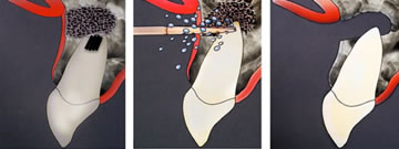
Lipoma
submandibular area / lateral surface of her neck. / cheek
the neoplasm has homogeneous, soft, and elastic consistency dough-like
mobile, and painless
not discolored
not fused with the skin or surrounding tissues
Its slow growth was observed for 3 years. 5 years
Infiltration anesthesia
anesthesia + lipoma
infiltration des lipoma et le median caries dans petit porcelain bridge
Fibroma
lateral surface of her tongue
s pink, mobile, painless, hard, and spherical
slowly growing
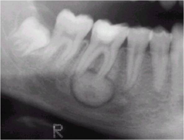
Odontoma
Orthopantomogram / xrayshows a
The density of the shadow is close to that of dental tissues / dentate shape and look like hard tooth tissues
On its periphery, the neoplasm is surrounded by a transparent zone 1 mm wide
… shadow + dental or dental + hard tooth tissues
Cementoma
The neoplasm is similar in its structure to the tooth root tissues
In the area of the root apex of tooth 45, a homogeneous round dense shadow
… homogenous wbhal bhal m3a l2rd
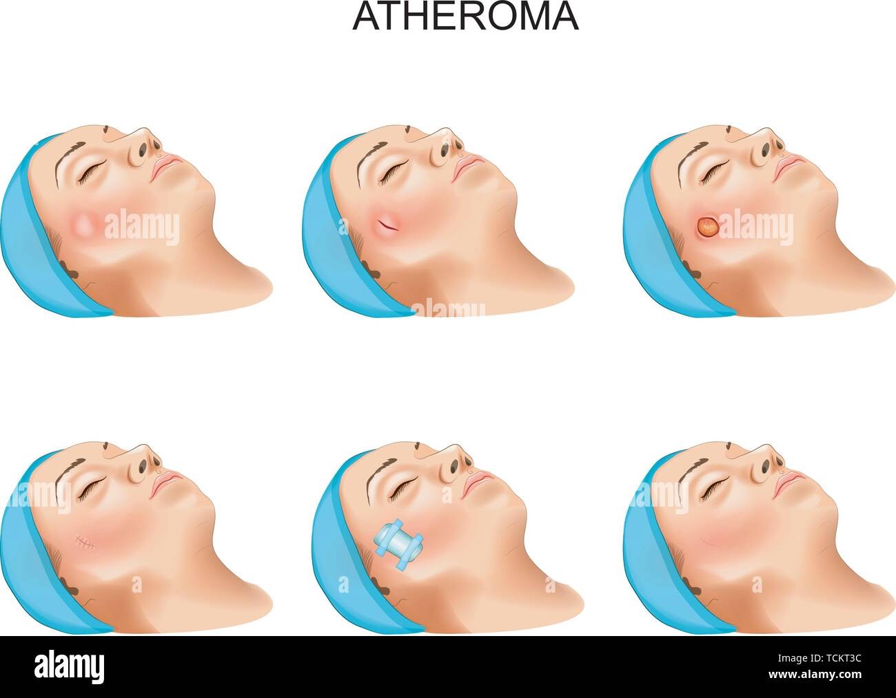
Atheroma
athe roma
slowly growing neoplasm
in the frontal area on the left / cheek
mobile, painless, and fused with the skin.
… fused to skin (2rdo) in frontal area slow in fight
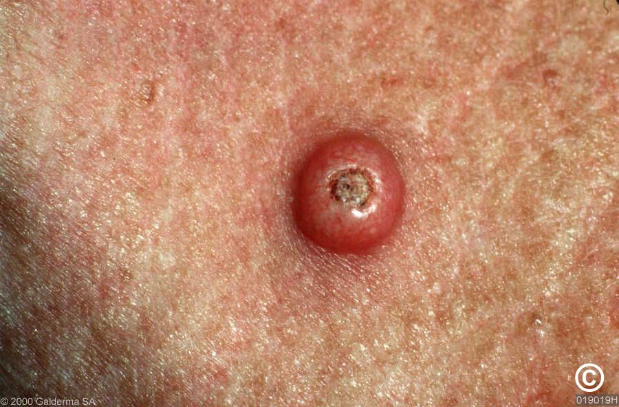
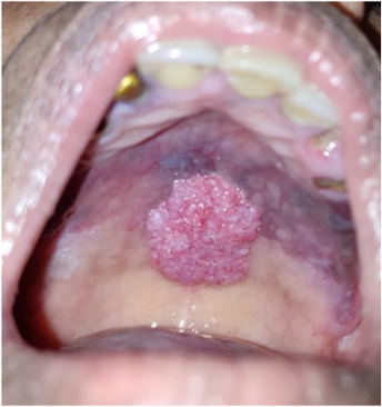
Keratoacanthoma
growing rapidly
gray-red nodule
filled with a dense keratinized mass / corneous masses
funnel shaped / with raised rolled edges / crater-like depression
Adenocarcinoma
dull pain in her left parotid-masseteric region
No saliva could be produced from the duct of the left parotid gland
paresis
adnan paresis w parotid masteric
granuloma / Migrating facial granuloma
infiltrate is sharply thinned-out, cyanotic, and glossy. On the oral mucosa along the mucogingival fold
right buccal area infiltration there is a fistula + destroyed by 2/3 with the fistula can be palpated
Odontogenic granuloma of the face
granuloma / Migrating facial granuloma + painless and slowly grows in size + dense band going from the growth to the destroyed 33rd tooth / Intraoral palpation reveals a cord going from the 37 tooth to the cheek lesion
Cystogranuloma
circular well-defined area of bone tissue destruction 0,7х 0,7 cm large in the projection of root apex
Granuloma removal with root apex resection
12 tooth is filled to the apex + granuloma 4 mm in diameter surrounds the root apex
treatment
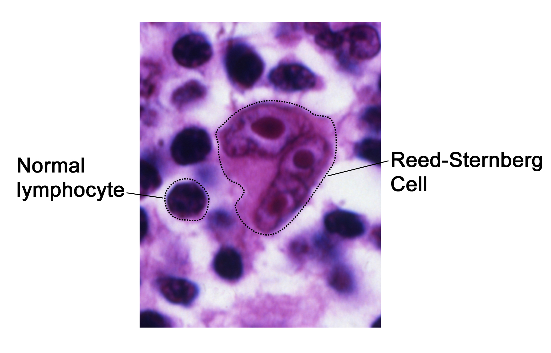
Lymphogranulomatosis
giant Reed-Sternberg cells
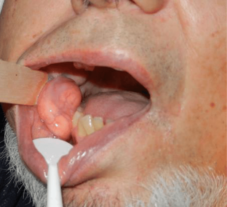
Angiomatous epulis
angi matou
bright-red soft painless formation that bleeds when palpated
an nssylhom dem
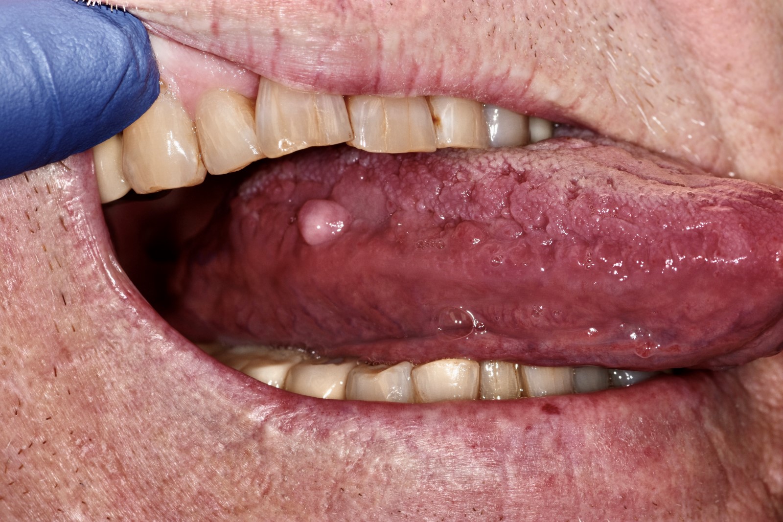
papilloma
les papillons
wart-like pedunculated growth / wide pedicle on the tongue apex / thin pedicle
or
The neoplasm sometimes increases, and sometimes decreases in size.
dans les pedicule (pedunculated ) parfois augmentent parfois non
Remove papillomas and make a denture with a double layered base lined with elastic material
needs a removable full laminar denture + the denture bed there are numerous dense papillomas
Prevention of malignant transformation of the papilloma. Removal of the papilloma
diagnosed with papilloma + tactics dental surgeon choose
Traumatic neuroma
The first complaints developed after a surgery in this area
The nodule is dense, elastic,
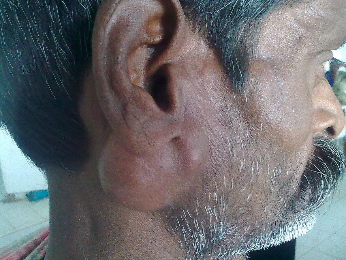
Madelung's deformity
made
excessive accumulation of adipose tissue in the patient's neck and upper torso
neck … by adipose
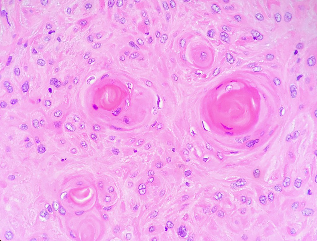
Bowen disease
keratin pearls are detected.
keratin ch3rk bah ouah
Giant-cell epulis
giant etpulis
a painless, tuberous, cyanotic pedunculated formation 2х1х1,5 cm large that appeared on the site of the extracted 46 tooth.
X-ray picture bone tissue destruction
tuberous pede est … detruit les os
cyst / Inborn median cyst
Puncture has yielded a yellow liquid with cholesterol crystals
Preliminary diagnosis of … = Yellowish liquid
types of cyst
+ anterior margin of the sternocleidomastoid = Lateral cervical cyst
+ the front surface of her neck on the midline = Midline cervical cyst
… + in the area of tooth 36 that was extracted 7 months ago =Residual cyst
absence of the periodontal fissure at the the palatal root apex / the periodontal fissure at the root apex is missing. orX-ray shows an area of bone destruction or A puncture of the neoplasm yielded a yellowish liquid with cholesterol crystals. = radicular cyst
Teeth 11 and 21 are intact. X-ray shows a homogeneous focus of bone tissue rarefaction = Nasopalatine duct cyst
44th tooth follicle is shifted down in distal direction = Follicular cyst of mandibula from the 44th tooth
bone thickening = Follicular cyst
there is a roundish, elastic, painless neoplasm / clear bluish content is visible through its walls + inside the lower lip. = Retention cyst of lower lip
roundish bulge up to 3 cm in diameter in the right sublingual or clear margins and signs of fluctuation+ The mucosa around this formation is unchanged = Retention cyst of the right sublingual salivary gland
Yellowish liquid
Preliminary diagnosis of maxillary radicular cyst was made
Lateral cervical cyst
Puncture has yielded a yellow liquid with cholesterol crystals + anterior margin of the sternocleidomastoid
Midline cervical cyst
Puncture has yielded a yellow liquid with cholesterol crystals + the front surface of her neck on the midline
Residual cyst
Puncture has yielded a yellow liquid with cholesterol crystals+ in the area of tooth 36 that was extracted 7 months ago
radicular cyst
absence of the periodontal fissure at the the palatal root apex / the periodontal fissure at the root apex is missing.
or
X-ray shows an area of bone destruction
radi abscence of periodontal fissure
Nasopalatine duct cyst / radicular cyst ( no naso )
bone tissue rarefaction
Follicular cyst / Follicular cyst of mandibula from the 44th tooth
bone thickening
44th tooth follicle is shifted down in distal direction
Retention cyst of lower lip / Retention cyst of the right sublingual salivary gland
there is a roundish, elastic, painless neoplasm / clear bluish content is visible through its walls + inside the lower lip.
.
roundish bulge up to 3 cm in diameter in the right sublingual or clear margins and signs of fluctuation + The mucosa around this formation is unchanged
Manganotti’s abrasive precancerous cheilitis
man
recurrent erosion
vermilion border of his lower lip
oval erosion / irregular shape
deep red color / glossy
covered in a scab
is deep glossy red recurrent in vermillon border of tinder
Verrucous precancerous lesion of the vermilion border
semicircle / hemispherical neoplasm neoplasm 1 cm
or
rounded tumor-like formation+covered with thin scales
Limited precancerous hyperkeratosis / Local precancerous hyperkeratosis of the lower lip / Localized precancerous hyperkeratosis
the lip midline and the mouth angle, there is a grayish-white irregular (polygonal) / the vermilion border of the lower lip, to the left from its center, there is a clearly demarkated polygonal
or
tumor-like growth on the red border of the left lower lip
tightly attached small scales
Surgical removal of the focus within healthy tissues
diagnosed with precancerous hyperkeratosis of the red border of the lower lip
Cancer of the tongue lateral surface / Cancer of the lateral surface of the tongue
complains of an ulcer on the lateral surface of the tongue
Despite these measures the ulcer continues to grow.
Lower lip cancer
condyloma on his lower lip
the red border of the lower lip is cyanotic and infiltrated,
Combined treatment
treatment + recurrence of lower lip cancer
Mandibular osteoradionecrosis
combined treatment for oral
mucosa cancer stage II (radiation therapy and surgery).
Cancer of the mucous membrane
He is smoker
12 years. of denture
mucous membrane in form of cauliflower,
Giant cell tumor of the mandible
bone destruction in the mandibular body
Tumor puncture yielded brown liquid
Malignant tumor of a parotid salivary gland
edema of the right parotid-masticatory region
When the patient tries to puff up his cheeks, the right cheek wavers
Ranula
ranula
neoplasm in her sublingual area
the neoplasm and is blue-tinged and semi-transparent
blue sublingual
Osteoclastoma
osteoclass
Puncture consists of brown exudate without cholesterol crystals.
Puncture consists of brown fluid without cholesterol crystals
l3kss dyal cyst
Marginal mandibular branch of facial nerve
Postoperatively benign tumor of the parotid gland
speech disturbance.
facial nerve was damaged during the surgical intervention?
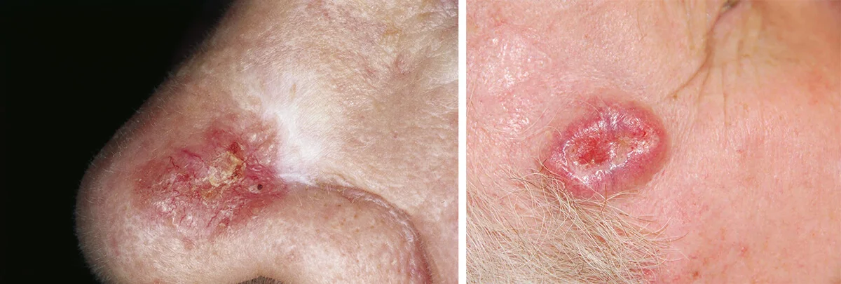
Basalioma
basal
ulcer on the right nasolabial sulcus
The ulcer grows in size and depth
nasolabial sulcus kbir fla forme wl 3om9 tikhrj mno …
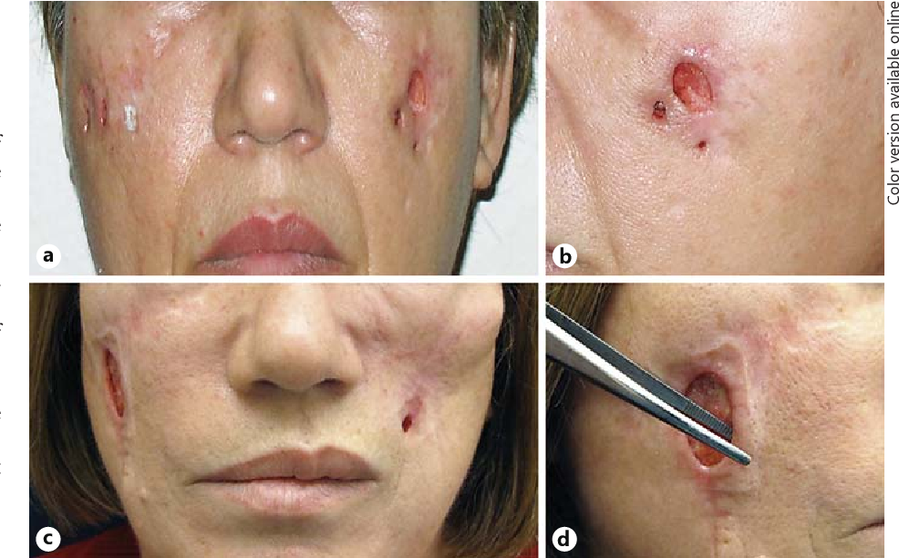
Mandibular resection at the distance of 1.5 cm from the lesion focus
surgery + diagnosed with mandibular ameloblastoma
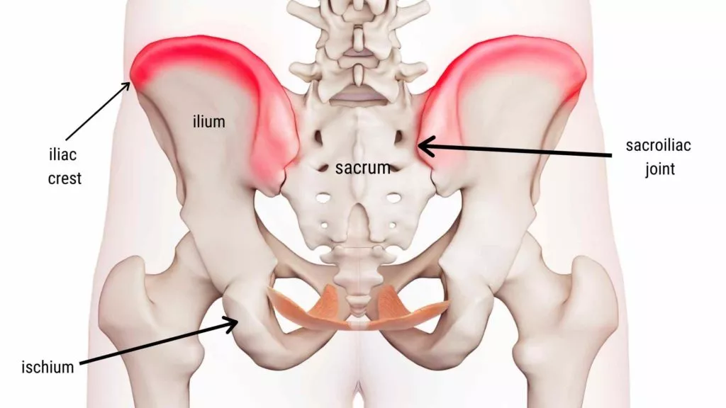
Iliac bone ridge
mandibular ameloblastoma
take a bone graft from ;